. 2009 Mar;10(1):21-6.
doi: 10.1007/s10195-008-0041-3. Epub 2009 Feb 3.

Cervical degenerative index: a new quantitative radiographic scoring system for cervical spondylosis with interobserver and intraobserver reliability testing

Affiliations

Cervical degenerative index: a new quantitative radiographic scoring system for cervical spondylosis with interobserver and intraobserver reliability testing

Elisha Ofiram et al. J Orthop Traumatol. 2009 Mar.

Abstract

Background: The lack of a widely available scoring system for cervical degenerative spondylosis encouraged the authors to establish and validate a systematic quantitative radiographic index.

Materials and methods: This study included intraobserver and interobserver reliability testing among three reviewers with different years of experience. Each observer independently scored four cervical radiographs of 48 patients at separate intervals, and statistical analysis of the grading was performed.

Results: There was high intraobserver and interobserver reliability between the two experienced observers. There was fair reliability between the less experienced observer and the more experienced observers.

Conclusions: The cervical degenerative index appears to be a reliable and reproducible radiographic assessment of cervical spondylosis. The index will have direct applicability for longitudinal study of cervical spondylosis and may be clinically relevant as well.
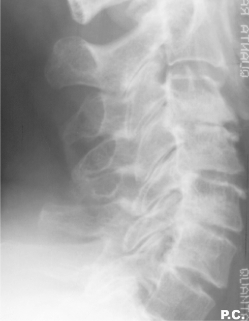
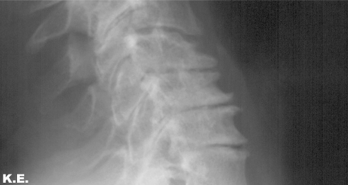
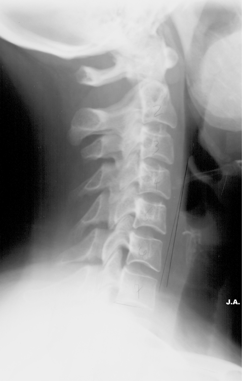

Figures

Fig. 1
Fig. 1
In this patient, we see the different grading scales for disc space narrowing rated by one observer as C2/3 = 0, C6/7 = 1, C4/5 = 2, C5/6 = 2, and C3/4 = 3
Fig. 2
Fig. 2
Severe endplate sclerosis is seen in a, and severe sclerosis of the facets at C2/3 without severe endplate sclerosis in b. Both were scored as 3 points
Fig. 3
Fig. 3
This shows osteophytes posteriorly, greater than 4 mm, yielding a score of 3. Also note the smaller anterior osteophytes
Fig. 4
Fig. 4
Flexion view showing 3–5 mm of subluxation yielding a factor score of 2 for that level
Fig. 5
Fig. 5
This patient had a scoliosis fusion from T2 to the sacrum at age 29, 8 years previously. Her lateral radiograph shows minimal degeneration (CDI 5)

References

    1. Kellgren JH, Ball J (1963) Atlas of standard radiographics, vol II. Blackwell Scientific, Oxford
    1. {'text': '', 'ref_index': 1, 'ids': [{'type': 'DOI', 'value': '10.1136/ard.17.4.388', 'is_inner': False, 'url': 'https://doi.org/10.1136/ard.17.4.388'}, {'type': 'PMC', 'value': 'PMC1007067', 'is_inner': False, 'url': 'https://pmc.ncbi.nlm.nih.gov/articles/PMC1007067/'}, {'type': 'PubMed', 'value': '13606727', 'is_inner': True, 'url': 'https://pubmed.ncbi.nlm.nih.gov/13606727/'}]}
    2. Kellgren JH, Lawrence JS (1958) Osteo-arthrosis and disk degeneration in an urban population. Ann Rheum Dis 17(4):388–397 - PMC - PubMed
    1. {'text': '', 'ref_index': 1, 'ids': [{'type': 'DOI', 'value': '10.1097/00007632-199704150-00007', 'is_inner': False, 'url': 'https://doi.org/10.1097/00007632-199704150-00007'}, {'type': 'PubMed', 'value': '9127918', 'is_inner': True, 'url': 'https://pubmed.ncbi.nlm.nih.gov/9127918/'}]}
    2. Cote P, Cassidy JD, Yong-Hing K, Sibley J, Loewy J (1997) Apophysial joint degeneration, disc degeneration, and sagittal curve of the cervical spine. Can they be measured reliably on radiographs? Spine 22(8):859–864 - PubMed
    1. {'text': '', 'ref_index': 1, 'ids': [{'type': 'DOI', 'value': '10.1097/00007632-198607000-00003', 'is_inner': False, 'url': 'https://doi.org/10.1097/00007632-198607000-00003'}, {'type': 'PubMed', 'value': '3787320', 'is_inner': True, 'url': 'https://pubmed.ncbi.nlm.nih.gov/3787320/'}]}
    2. Gore DR, Sepic SB, Gardner GM (1986) Roentgenographic findings of the cervical spine in asymptomatic people. Spine 11(6):521–524 - PubMed
    1. Ofiram E, Garvey TA, Schwender JD, Wroblewski JM, Winter RB (2009) Cervical degenerative changes in idiopathic scoliosis patients who underwent long fusion to the sacrum as adults: incidence, severity, and evolution. J Orthopaed Traumatol 10(1). doi: 10.1007/s10195-008-0044-0 - PMC - PubMed

LinkOut - more resources