

Modulation of preparatory volitional motor cortical activity by paired associative transcranial magnetic stimulation
- PMID: 19384889
- PMCID: PMC6870586
- DOI: 10.1002/hbm.20793
Modulation of preparatory volitional motor cortical activity by paired associative transcranial magnetic stimulation
Abstract
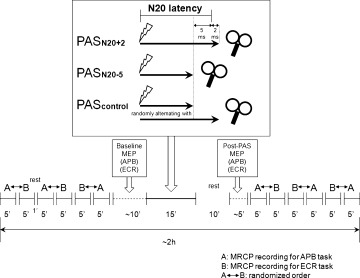
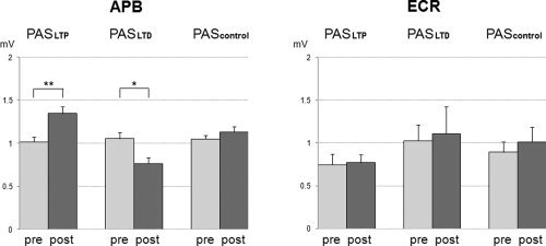
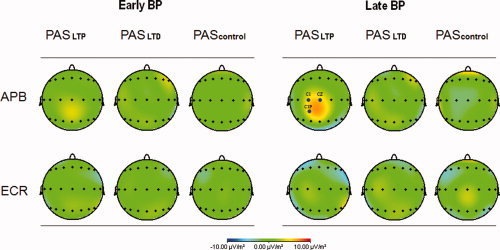
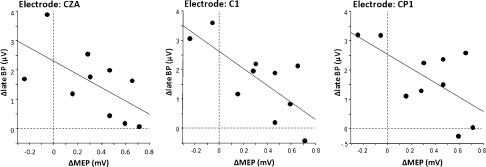
Paired associative transcranial magnetic stimulation (PAS) has been shown to induce long-term potentiation (LTP)-like or long-term depression (LTD)-like change in excitability of human primary motor cortex (M1), as probed by motor evoked potential (MEP) amplitude. In contrast, little is known about PAS effects on volitional motor cortical activity. In 10 healthy subjects, movement related cortical potentials (MRCP) were recorded to index volitional motor cortical activity during preparation of simple thumb abduction (prime mover: abductor pollicis brevis, APB) or wrist extension movements (prime mover: extensor carpi radialis, ECR). PAS(LTP) increased, PAS(LTD) decreased, and PAS(control) did not change MEP(APB), while MEP(ECR), not targeted by PAS, remained unchanged in all PAS conditions. PAS(LTP) decreased MRCP negativity during the late Bereitschaftspotential (-500 to 0 ms before movement onset), only in the APB task, and predominantly over central scalp electrodes contralateral to the thumb movements. This effect correlated negatively with the PAS(LTP) induced increase in MEP(APB). PAS(LTD) and PAS(control) did not affect MRCP amplitude. Findings indicate a specific interference of PAS with preparatory volitional motor cortical activity, suggestive of a net result caused by increased M1 excitability and disrupted effective connectivity between premotor areas and M1.
Figures

References
-
- Agostino R, Iezzi E, Dinapoli L, Gilio F, Conte A, Mari F, Berardelli A ( 2007): Effects of 5 Hz subthreshold magnetic stimulation of primary motor cortex on fast finger movements in normal subjects. Exp Brain Res 180: 105–111. - PubMed
-
- Caporale N, Dan Y ( 2008): Spike timing‐dependent plasticity: A Hebbian learning rule. Annu Rev Neurosci 31: 25–46. - PubMed
-
- Chen R, Classen J, Gerloff C, Celnik P, Wassermann EM, Hallett M, Cohen LG ( 1997): Depression of motor cortex excitability by low‐frequency transcranial magnetic stimulation. Neurology 48: 1398–1403. - PubMed
-
- Chen R, Hallett M ( 1999): The time course of changes in motor cortex excitability associated with voluntary movement. Can J Neurol Sci 26: 163–169. - PubMed
Publication types
MeSH terms
LinkOut - more resources
Full Text Sources
