Evaluation at atomic resolution of the role of strain in destabilizing the temperature-sensitive T4 lysozyme mutant Arg 96 --> His
- PMID: 19384984
- PMCID: PMC2771290
- DOI: 10.1002/pro.93
Evaluation at atomic resolution of the role of strain in destabilizing the temperature-sensitive T4 lysozyme mutant Arg 96 --> His
Abstract
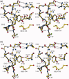
Mutant R96H is a classic temperature-sensitive mutant of bacteriophage T4 lysozyme. It was in fact the first variant of the protein to be characterized structurally. Subsequently, it has been studied extensively by a variety of experimental and computational techniques, but the reasons for the loss of stability of the mutant protein remain controversial. In the crystallographic refinement of the mutant structure at 1.9 A resolution one of the bond angles at the site of substitution appeared to be distorted by about 11( degrees ), and it was suggested that this steric strain was one of the major factors in destabilizing the mutant. Different computationally-derived models of the mutant structure, however, did not show such distortion. To determine the geometry at the site of mutation more reliably, we have extended the resolution of the data and refined the wildtype (WT) and mutant structures to be better than 1.1 A resolution. The high-resolution refinement of the structure of R96H does not support the bond angle distortion seen in the 1.9 A structure determination. At the same time, it does confirm other manifestations of strain seen previously including an unusual rotameric state for His96 with distorted hydrogen bonding. The rotamer strain has been estimated as about 0.8 kcal/mol, which is about 25% of the overall reduction in stability of the mutant. Because of concern that contacts from a neighboring molecule in the crystal might influence the geometry at the site of mutation we also constructed and analyzed supplemental mutant structures in which this crystal contact was eliminated. High-resolution refinement shows that the crystal contacts have essentially no effect on the conformation of Arg96 in WT or on His96 in the R96H mutant.
Figures


Similar articles
-
Contributions of all 20 amino acids at site 96 to the stability and structure of T4 lysozyme.Protein Sci. 2009 May;18(5):871-80. doi: 10.1002/pro.94. Protein Sci. 2009. PMID: 19384988 Free PMC article.
-
The introduction of strain and its effects on the structure and stability of T4 lysozyme.J Mol Biol. 2000 Jan 7;295(1):127-45. doi: 10.1006/jmbi.1999.3300. J Mol Biol. 2000. PMID: 10623513
-
Structure of oxidized bacteriophage T4 glutaredoxin (thioredoxin). Refinement of native and mutant proteins.J Mol Biol. 1992 Nov 20;228(2):596-618. doi: 10.1016/0022-2836(92)90844-a. J Mol Biol. 1992. PMID: 1453466
-
Lessons from the lysozyme of phage T4.Protein Sci. 2010 Apr;19(4):631-41. doi: 10.1002/pro.344. Protein Sci. 2010. PMID: 20095051 Free PMC article. Review.
-
The tail lysozyme complex of bacteriophage T4.Int J Biochem Cell Biol. 2003 Jan;35(1):16-21. doi: 10.1016/s1357-2725(02)00098-5. Int J Biochem Cell Biol. 2003. PMID: 12467643 Review.
Cited by
-
Interaction energy based protein structure networks.Biophys J. 2010 Dec 1;99(11):3704-15. doi: 10.1016/j.bpj.2010.08.079. Biophys J. 2010. PMID: 21112295 Free PMC article.
-
Metal ions-binding T4 lysozyme as an intramolecular protein purification tag compatible with X-ray crystallography.Protein Sci. 2017 Jun;26(6):1116-1123. doi: 10.1002/pro.3162. Epub 2017 Apr 2. Protein Sci. 2017. PMID: 28342173 Free PMC article.
-
Simplifying and enhancing the use of PyMOL with horizontal scripts.Protein Sci. 2016 Oct;25(10):1873-82. doi: 10.1002/pro.2996. Epub 2016 Aug 16. Protein Sci. 2016. PMID: 27488983 Free PMC article.
-
Large language models generate functional protein sequences across diverse families.Nat Biotechnol. 2023 Aug;41(8):1099-1106. doi: 10.1038/s41587-022-01618-2. Epub 2023 Jan 26. Nat Biotechnol. 2023. PMID: 36702895 Free PMC article.
-
Contributions of all 20 amino acids at site 96 to the stability and structure of T4 lysozyme.Protein Sci. 2009 May;18(5):871-80. doi: 10.1002/pro.94. Protein Sci. 2009. PMID: 19384988 Free PMC article.
References
-
- Grütter MG, Hawkes RB, Matthews BW. Molecular basis of thermostability in the lysozyme from bacteriophage T4. Nature. 1979;277:667–669. - PubMed
-
- Hawkes R, Grütter MG, Schellman J. Thermodynamic stability and point mutations of bacteriophage T4 lysozyme. J Mol Biol. 1984;175:195–212. - PubMed
-
- Becktel WJ, Baase WA. Thermal denaturation of bacteriophage T4 lysozyme at neutral pH. Biopolymers. 1987;26:619–623. - PubMed
-
- Becktel WJ, Schellman JA. Protein stability curves. Biopolymers. 1987;26:1859–1877. - PubMed
-
- Kitamura S, Sturtevant JM. A scanning calorimetric study of the thermal denaturation of the lysozyme of phage T4 and the Arg96→His mutant form thereof. Biochemistry. 1989;28:3788–3792. - PubMed
Publication types
MeSH terms
Substances
Grants and funding
LinkOut - more resources
Full Text Sources

