. 2009 May;18(5):921-35.
doi: 10.1002/pro.101.

Apolipoprotein AI tertiary structures determine stability and phospholipid-binding activity of discoidal high-density lipoprotein particles of different sizes

Affiliations

Apolipoprotein AI tertiary structures determine stability and phospholipid-binding activity of discoidal high-density lipoprotein particles of different sizes

Bin Chen et al. Protein Sci. 2009 May.

Abstract

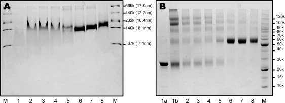
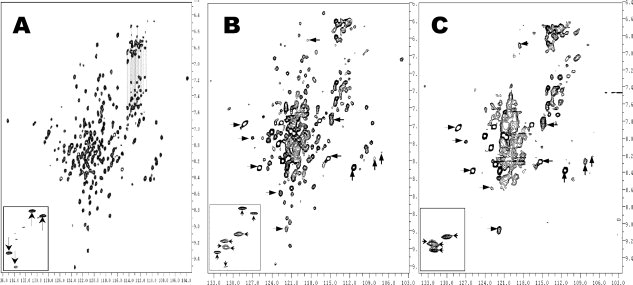
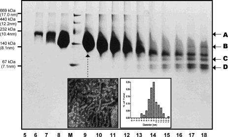
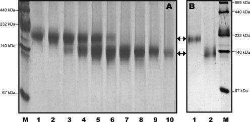
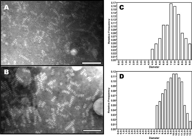
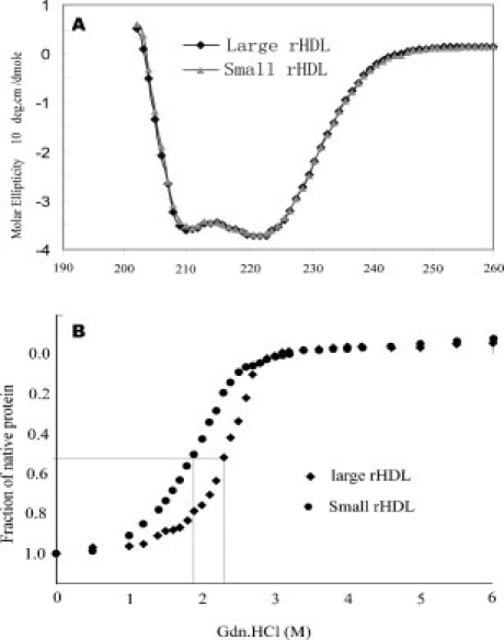
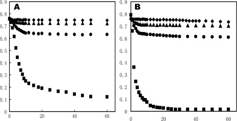
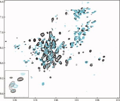
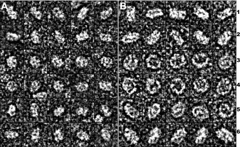
Human high-density lipoprotein (HDL) plays a key role in the reverse cholesterol transport pathway that delivers excess cholesterol back to the liver for clearance. In vivo, HDL particles vary in size, shape and biological function. The discoidal HDL is a 140-240 kDa, disk-shaped intermediate of mature HDL. During mature spherical HDL formation, discoidal HDLs play a key role in loading cholesterol ester onto the HDL particles by activating the enzyme, lecithin:cholesterol acyltransferase (LCAT). One of the major problems for high-resolution structural studies of discoidal HDL is the difficulty in obtaining pure and, foremost, homogenous sample. We demonstrate here that the commonly used cholate dialysis method for discoidal HDL preparation usually contains 5-10% lipid-poor apoAI that significantly interferes with the high-resolution structural analysis of discoidal HDL using biophysical methods. Using an ultracentrifugation method, we quickly removed lipid-poor apoAI. We also purified discoidal reconstituted HDL (rHDL) into two pure discoidal HDL species of different sizes that are amendable for high-resolution structural studies. A small rHDL has a diameter of 7.6 nm, and a large rHDL has a diameter of 9.8 nm. We show that these two different sizes of discoidal HDL particles display different stability and phospholipid-binding activity. Interestingly, these property/functional differences are independent from the apoAI alpha-helical secondary structure, but are determined by the tertiary structural difference of apoAI on different discoidal rHDL particles, as evidenced by two-dimensional NMR and negative stain electron microscopy data. Our result further provides the first high-resolution NMR data, demonstrating a promise of structural determination of discoidal HDL at atomic resolution using a combination of NMR and other biophysical techniques.


Figures

Figure 1
Figure 1
Panel A: A 6–20% gradient native PAGE gel of rHDL particles prepared using different POPC:ApoAI ratios (POPC/cholate = 0.737). Panel B: A 6–12% tricine gel of crosslinking results of rHDL particles using different POPC:ApoAI ratios. In both panels, lane 2: POPC:ApoAI = 5:1, lane 3: POPC:ApoAI = 10:1, lane 4: POPC:ApoAI = 20:1, lane 5: POPC:ApoAI = 40:1, lane 6: POPC:ApoAI = 60:1, lane 7: POPC:ApoAI = 80:1, lane 8: POPC:ApoAI = 100:1, lane M: molecular marker. In Panel A: lane 1: lipid-free apoAI. In Panel B: lane 1a: lipid-free apoAI without crosslinker, lane 1b: lipid-free apoAI with crosslinker.
Figure 2
Figure 2
[15N, H] TROSY-HSQC spectra of Lp1AI (Panel A), rHDL sample before the first ultracentrifugation (Panel B) and rHDL sample after the first ultracentrifugation (Panel C). These spectra were recorded on a Varian INOVA 500 MHz spectrometer with a triple-resonance room temperature probe. The insets of different panels show the crosspeaks of the Trp sidechain Hɛ1/Nɛ1 atoms. The NMR samples are in 50 mM phosphate buffer at pH7.0, with 50 mM NaCl, 5 mM EDTA, 0.5 mM NaN3, and 0.05 mM DSS.
Figure 3
Figure 3
A 4–20% native gel of the fractions of the first ultracentrifugation of an rHDL preparation. A total of 20 fractions have been collected and fractions 1–4 and 19–20 contain no apoAI proteins. Fraction 1 is the top fraction and fraction 20 is the bottom fraction. M: High molecular weight marker. Arrows A and B show the broad range of the purified discoidal rHDL. Arrows C and D show several fractions of lipid-poor apoAI. Inset: Negative stain electron microscopic images of rHDL particles of Lane 9 (Left) and the size distribution determined by electron microscopy (Right). The average size of the discoidal HDL: 8.8 ± 3.7 nm.
Figure 4
Figure 4
Panel A: A 4–20% native gel of the fractions of the last ultracentrifugation, showing the purification of small and large discoidal rHDL particles can be achieved using this ultracentrifugation method. Panel B: A 4–20% native gel of the purified large (Lane 1) and small (Lane 2) discoidal rHDL. M: High molecular weight marker.
Figure 5
Figure 5
Panel A: PTA negative stain electron microscopy of purified small discoidal rHDL. Panel B: Negative stain electron microscopy of the purified large discoidal rHDL. Bar = 50 nm. Panel C: Size distribution of purified small discoidal rHDL. Panel D: Size distribution of purified large discoidal rHDL. The average diameter of small discoidal rHDL is 7.6 ± 1.6 nm and average diameter of large discoidal rHDL is: 9.8 ± 1.7 nm.
Figure 6
Figure 6
Panel A: Far-UV CD spectra of small (Black) and large discoidal rHDL (Gray) at 0.2 mg/mL in 50 mM sodium phosphate, 150 mM NaCl, pH 7.2. Panel B: GdnHCl denaturation of small (Circle) and large discoidal rHDL (diamond) under the same condition. The fraction of unfolding was plotted as a function of GndHCl concentration.
Figure 7
Figure 7
Phospholipid vesicle clearance. DMPC vesicles were incubated at 23.9°C in the absence (diamond) and presence of lipid-free apoAI (square), large rHDL (triangle) and small rHDL (Circle) in 20 mM Tris-HCl, pH 7.2, 250 mM NaCl, and 1 mM EDTA. Vesicle clearance as a function of time was followed by OD at 490 nm. Panel A: DMPC to apoAI (w/w) is 2:1 and Panel B: DMPC to apoAI (w/w) is 1:1.
Figure 8
Figure 8
A comparison of [15N, 1H] TROSY-HSQC spectra of small rHDL (black) and large rHDL (Blue) collected on a 600 MHz NMR instrument with a cold probe at 30°C. NMR samples contain 0.2–0.5 mM triple-labeled rHDL in 50 mM phosphate buffer containing 50 mM NaCl, 0.1 mM NaN3, 1 mM EDTA, 5–7% D2O at pH 7.2. Inset: the crosspeaks of Trp sidechain Hɛ1/Nɛ1 atoms of the apoAI on small (Black) and large (Blue) discoidal rHDL particles.
Figure 9
Figure 9
Uranyl formate negatively stained electron microscopy of purified small and large discoidal rHDLs. Panel A displays 30 representative small rHDL particle and Panel B displays 30 representative large rHDL particles. Rows 1 and 2 are the side view; Rows 3–4 are the top view; Rows 5–6 are the tilted top view of both small (A) and large (B) rHDL particles. Each box corresponds to a 15 × 15 nm area.

Similar articles

Cited by

References

    1. Lewis GF, Rader DJ. New insights into the regulation of HDL metabolism and reverse cholesterol transport. Circ Res. 2005;96:1221–1232. - PubMed
    1. Davidson WS, Thompson TB. The structure of apolipoprotein a-I in high-density lipoproteins. J Biol Chem. 2007;282:22249–22253. - PubMed
    1. Asztalos BF. High-density lipoprotein metabolism and progression of atherosclerosis: new insights from the HDL Atherosclerosis Treatment Study. Curr Opin Cardiol. 2004;19:385–391. - PubMed
    1. Duriez P, Fruchart JC. High-density lipoprotein subclasses and apolipoprotein A-I. Clin Chim Acta. 1999;286:97–114. - PubMed
    1. Martinez LO, Jacquet S, Terce F, Collet X, Perret B, Barbaras R. New insight on the molecular mechanisms of high-density lipoprotein cellular interactions. Cell Mol Life Sci. 2004;61:2343–2360. - PMC - PubMed

Publication types

MeSH terms

LinkOut - more resources