. 2009 May;18(5):960-9.
doi: 10.1002/pro.106.

Nanoscale elongating control of the self-assembled protein filament with the cysteine-introduced building blocks

Affiliations

Nanoscale elongating control of the self-assembled protein filament with the cysteine-introduced building blocks

Kengo Usui et al. Protein Sci. 2009 May.

Erratum in

  • Protein Sci. 2009 Jul;18(7):1571

Abstract

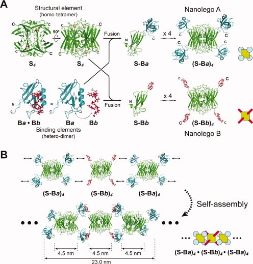
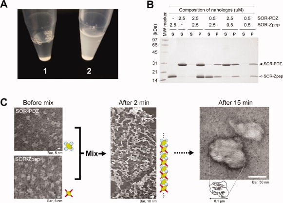
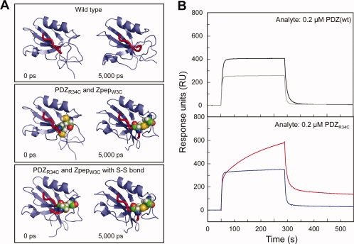
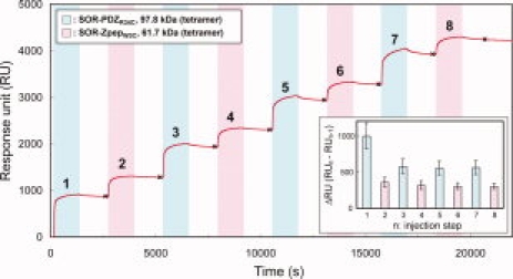
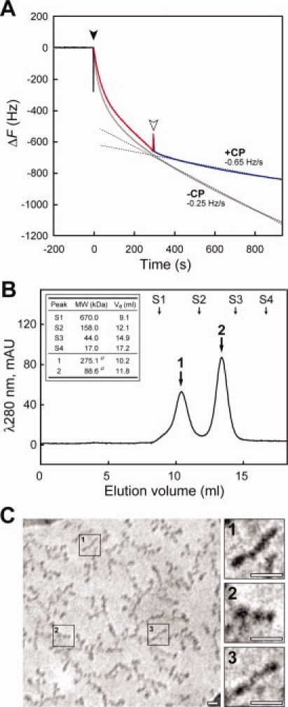
Self-assembly of artificially designed proteins is extremely desirable for nanomaterials. Here we show a novel strategy for the creation of self-assembling proteins, named "Nanolego." Nanolego consists of "structural elements" of a structurally stable symmetrical homo-oligomeric protein and "binding elements," which are multiple heterointeraction proteins with relatively weak affinity. We have established two key technologies for Nanolego, a stabilization method and a method for terminating the self-assembly process. The stabilization method is mediated by disulfide bonds between Cysteine-residues incorporated into the binding elements, and the termination method uses "capping Nanolegos," in which some of the binding elements in the Nanolego are absent for the self-assembled ends. With these technologies, we successfully constructed timing-controlled and size-regulated filament-shape complexes via Nanolego self-assembly. The Nanolego concept and these technologies should pave the way for regulated nanoarchitecture using designed proteins.


Figures

Figure 1
Figure 1
Design of self-assembling Nanolegos. A: Construction strategy of two hetero-Nanolegos, SOR-PDZ and SOR-Zpep. The basic crystal structure, indicated as structural (S) and binding elements (Ba and Bb), was composed of P. furiosus SOR and human Erbin PDZ domain and the binding peptide, respectively. For modeling of tetrameric SOR-PDZ and SOR-Zpep fusion proteins, we first constructed the model structure of monomeric SOR-PDZ and SOR-Zpep fusion proteins. The models of tetrameric fusion proteins, (S-Ba)4 and (S-Bb)4, were drawn by reconfiguration of the tetramer at the part of SOR. B: Putative self-assembling pattern of filamentous Nanolegos. These models were obtained by MD simulation based on the putative structure of the tetramer Nanolego in Figure 1(A) and reconfiguration of PDZ/Zpep-complex.
Figure 2
Figure 2
SOR Nanolegos self-assembling to the filamentous form. A: Change of state in bulk-solution by mixing purified SOR-PDZ and SOR-Zpep. The final concentration of each Nanolego was 10 μM in mixed solution. Each purified Nanolego formed a clear solution as shown in Photo 1. Photo 2 shows the solution state after 20 min from mixing these Nanolegos. B: SDS-PAGE analysis for the turbidity of SOR Nanolego mixture. The turbidity appeared in two Nanolegos-mixed solution was separated as the insoluble fraction by centrifugation at 14000g for 30 min at 4°C. The soluble and insoluble fractions of the mixture were subjected to SDS-PAGE in the lanes indicated by “S” and “P,” respectively. Each Nanolego was observed as single band with the mobility of monomer, 28.2 kDa (SOR-PDZ) and 15.7 kDa (SOR-Zpep) under the denatured condition with SDS. C: Negatively stained transmission electron micrograph of the self-assembled Nanolegos. As a rough approximation, the diameter of the filamentous form (after 2 min) was about 5 nm.
Figure 3
Figure 3
Construction of Cys-introduced Nanolegos. A: Introduction of a disulfide bond to between PDZ domain and PDZ-binding peptide. The results were MD simulation of the interaction between PDZR34C and ZpepW3C mutant. The structures on the left side are initial models before MD (0 ps), and those on the right side are the final models after MD for 5000 ps. B: Binding analyses between PDZ- and Zpep-Cys-mutant by the SPR assay. All sensorgrams indicate the response when 0.2 μM analyte was used. The kinetics values of each binding assay are shown in Supporting Information Table 2.
Figure 4
Figure 4
The sensorgram of 8-steps extension assembly of Cys-introduced Nanolegos. At every injection step, SOR-PDZR34C (colored sky-blue) or SOR-ZpepW3C (colored pink) was loaded for 20 min. The washing steps (not colored) were performed by passing the running buffer for 20 min. The inset graph is the summary of the increment of the SPR response for each injection of Nanolegos. The difference of RU at each injection step (ΔRU = RUn− RUn−1; n, injection step) was calculated from the RU at the data points shown as “×” in this figure. This graph presents the results of triplicate analyses, and the error bar on each column shows the standard deviation of each injection step.
Figure 5
Figure 5
Effect of CP Nanolego on extension of Nanolego filament. A: Terminating effect of CP-SOR-ZpepW3C on filament extension observed by QCM assay for a batch solution. Black and white arrowheads indicate the injection point of SOR-PDZR34C (at 0 s) and CP-SOR-ZpepW3C (at 300 s), respectively. The main sensorgram to investigate the effect of CP-SOR-ZpepW3C is colored separately in three sections; baseline (black, −200 to –0 s), filament extension with two Nanolegos mixture (red, 0–300 s), and addition of CP-SOR-ZpepW3C (blue, 300–700 s). The gray sensorgram shows the result without the addition of CP-SOR-ZpepW3C as the control experiment. The dashed lines are the result of approximated curve fitting of the sensorgram with or without the addition of CP-SOR-ZpepW3C, and each slope of the curve as the frequency change rate is indicated in the graph with “+CP” or “−CP”, respectively. B: The result of gel-filtration chromatography of the mixture of both SOR-PDZR34C and CP-SOR-ZpepW3C in solution. The mixture of two Nanolegos was performed by the concentration ratio of SOR-PDZR34C:CP-SOR-ZpepW3C = 1:5. Peak a and b corresponded to the 3-mer Nanolego complex ([CP-SOR-ZpepW3C] × 2 + [SOR-PDZR34C] × 1) and single CP-SOR-ZpepW3C, respectively. Arrows marked with S1–4 show the eluting volumes of the molecular standard proteins. Details of elution peak a, b and the standard proteins are shown in the inset table. “Ve” indicates each elution volume. a, theoretical molecular weight. C: The results of CryoTEM observation of putative 3-mer Nanolego complexes. Photographs on the right are threefold magnifications of the three areas on the left indicated by numbers. Each white bar indicates a scale of 10 nm.

References

    1. Whitesides GM, Boncheva M. Beyond molecules: self-assembly of mesoscopic and macroscopic components. Proc Natl Acad Sci USA. 2002;99:4769–4774. - PMC - PubMed
    1. Zhang S. Fabrication of novel biomaterials through molecular self-assembly. Nat Biotechnol. 2003;21:1171–1178. - PubMed
    1. Padilla JE, Colovos C, Yeates TO. Nanohedra: using symmetry to design self assembling protein cages, layers, crystals, and filaments. Proc Natl Acad Sci USA. 2001;98:2217–2221. - PMC - PubMed
    1. Yeates TO, Padilla JE. Designing supramolecular protein assemblies. Curr Opin Struct Biol. 2002;12:464–470. - PubMed
    1. Ringler P, Schulz GE. Self-assembly of proteins into designed networks. Science. 2003;302:106–109. - PubMed

Publication types

MeSH terms

LinkOut - more resources