Review
. 2009 Apr;88(4):729-37.
doi: 10.1016/j.exer.2009.01.015.

The washout phenomenon in aqueous outflow--why does it matter?

Affiliations
Review

The washout phenomenon in aqueous outflow--why does it matter?

Haiyan Gong et al. Exp Eye Res. 2009 Apr.

Abstract

The washout effect is a phenomenon in which the resistance to aqueous outflow diminishes with the volume of perfusate flowing through the outflow pathways, even if the perfusate is aqueous humor itself. One intriguing aspect of this phenomenon is that it appears to occur in the eyes of all species studied to date except humans. Even non-human primate eyes exhibit washout.

Because washout does not occur in human eyes some have concluded that a greater understanding of this effect could not be relevant to the study of human primary open angle glaucoma. Those who have chosen to study this phenomenon realize that if a washout effect could be induced in the human eye, the result would be a reduction in outflow resistance and a drop in intraocular pressure – precisely the goal of all current therapy for open angle glaucoma.

This article reviews the discovery of this phenomenon, the various lines of investigation aimed at unraveling its underlying mechanisms. It concludes with recent structural and functional comparisons that point to clear differences in the connectivity between the inner wall (IW) endothelial cells of Schlemm’s canal and matrix or cells in the juxtacanalicular connective tissue (JCT) between human eyes that do not exhibit washout and non-human eyes that do exhibit washout. This enhanced connectivity consisted of a more complex array of elastic fiber connections between the IW and JCT in human eyes. This enhanced connectivity may withstand the hydrodynamic forces driving separation between the IW and JCT, which occurs in non-human eyes during washout. Strategies targeting JCT/IW or JCT/JCT connectivity in human eyes might be promising anti-glaucoma therapies to decrease outflow resistance, and thus IOP.


Figures

Figure 1
Figure 1. “Washout” in bovine eyes but not in humans
Anterior segments from human eyes (n = 13) and neonatal calf eyes (n= 9) were perfused at 15 mmHg with Dulbecco’s modified eagle medium (DMEM) in a 5% CO2 environment at 37°C. A progressive increase in facility of outflow over baseline facility (%C0) with time was noted in the calf eyes but not in the human eyes. Erickson-Lamy et al: Absence of time-dependent facility increase (“washout”) in the perfused enucleated human eye. Invest Ophthalmol Vis Sci 31:2384–2388, 1990
Figure 2
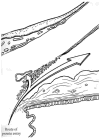
Figure 2. The principal route of entry for plasma-derived proteins into the aqueous humor of the normal eye
Note the route of passage delivers protein in close proximity to the trabecular meshwork and aqueous outflow pathway. Morrison, J. C. and Freddo, T. F. (1996). Anatomy, microcirculation and ultrastructure of the ciliary body. In: The Glaucomas. 2nd ed. Ritch, R., Shields, B and Krupin, T., eds., Mosby/W.B. Saunders, St. Louis, MO., U.S.A., Cptr. 6, p. 137.
Figure 3
Figure 3. Morphologic comparison between long- and short-duration perfusion in bovine eyes
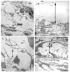
A. Transmission Electron Micrograph of Bovine Aqueous Plexus and underlying JCT region after Long-Duration Perfusion Significant separation between the basal lamina (BL) of the inner wall (IW) of the bovine aqueous plexus (AP) and underlying extracellular matrix of the JCT region is observed (double headed arrow). Scale bar: 5μm B. Transmission Electron Micrograph of Bovine Aqueous Plexus and underlying JCT region after Short-Duration Perfusion No distention of the JCT or separation of the IW lining of the aqueous plexus (AP) was observed. Giant vacuoles (GV) were seen along the inner wall. Scale bar: 5μm (Scott PA, Overby DR, Freddo TF, Gong H: Comparative studies between species that do and do not exhibit the washout effect. Exp. Eye Res, 84:435–443, 2007)
Figure 4
Figure 4. Correlation of Absolute Value of Pre-Fixation Facility and JCT/Inner Wall Separation Scores
The absolute value of outflow facility measured prior to fixation correlated with the extent of inner wall/JCT separation in all bovine eyes (p=0.0024). Closed circles = long-duration, open circles = short-duration. (Scott PA, Overby DR, Freddo TF, Gong H: Comparative studies between species that do and do not exhibit the washout effect. Exp. Eye Res, 84:435–443, 2007.)
Figure 5
Figure 5. An illustration of the streamlines of aqueous flow through the JCT before (A) and after (B) washout, as predicted by the funneling hypothesis
A. To enter Schlemm’s canal, aqueous must converge to pass through discrete openings along the inner wall, which decreases the available area for flow and increases outflow resistance. B. Separation of the inner wall from the JCT eliminates the funneling pattern, increases the available area for flow, and increases outflow facility during washout. (Overby D, Gong H, Freddo TF and Johnson M: The mechanism of increasing outflow facility during washout in the bovine eye. Invest Ophthalmol Vis Sci. 43:3455–3464, 2002.)
Figure 6
Figure 6. Morphologic change in monkey eyes during washout
Separation between the JCT cells and the JCT cells and matrix were often observed (double arrows). This micrograph was from a tracer study, microspheres were seen in the JCT (*). (From Dr. Haiyan Gong)
Figure 7
Figure 7. Morphologic comparison between long and short during perfusion in human eyes
A. Transmission electron micrograph of human Schlemm’s canal and underlying JCT region after short-duration perfusion. B. Transmission electron micrograph of human Schlemm’s canal and underlying JCT region after long-duration perfusion. No separation is observed between the inner wall (IW) of Schlemm’s canal (SC) and the underlying JCT in both short- and long-duration perfusion (A and B, respectively). Giant vacuoles (GV) are seen along the IW of SC with a similar distribution in both short and long-duration perfusion. Compared to bovine eyes (Fig. 3A and B) there is a more complex array of elastic fibers (EL) in the JCT region (single headed arrows), especially in the area immediately underneath the inner wall (IW). Scale bars: 3 mm. (Scott PA, Overby DR, Freddo TF, Gong H: Comparative studies between species that do and do not exhibit the washout effect. Exp. Eye Res, 84:435–443, 2007.)
Figure 8
Figure 8. Morphological comparison between washout effect and the effect of certain drugs that disrupt the cytoskeleton
Similar morphological changes were observed between washout (A) and Y27632 treatment (B) in bovine eyes and between washout (C) and latrunculin-B treatment (D) in monkey eyes. In A and B, separations were seen between the inner wall and JCT (double arrow). In C and D, separations between the cell-cell and cell and matrix were seen in the JCT regions (double arrow). SC, Schlemm’s canal; IW, inner wall; JXT (JCT), juxtacanalicular region; OW, outer wall; P, SUB, sub-canalicular cells (A from Overby D, Gong H, Freddo TF and Johnson M: The mechanism of increasing outflow facility during washout in the bovine eye. Invest Ophthalmol Vis Sci. 43:3455–3464, 2002; B from Lu Z, Overby DR, Scott PA, Freddo TF, Gong H: The mechanism of increasing outflow facility by rho-kinase inhibition with Y-27632 in bovine eyes. Exp Eye Res, 86: 271–281, 2008; C from Dr. Haiyan Gong; and D from Sabanay, I., B. Tian, B.T. Gabelt, B. Geiger, and P.L. Kaufman. 2006. Latrunculin B effects on trabecular meshwork and corneal endothelial morphology in monkeys. Exp Eye Res. 82:236–46).
Figure 9
Figure 9. Confocal (A, B) and Light microscopic (C, D) analysis
A. In a control eye, a segmental distribution of tracers (pink color) was seen along the inner wall of aqueous plexus (AP). B. In an Y27632-treated eye, the tracer distribution (pink color) was more uniform along the inner wall of AP. C. No separation was found between the inner wall of aqueous plexus (AP) and juxtacanalicular connective tissue (JCT) in a control eye. D. Separation was found between the inner wall of AP and JCT (double arrow) in an Y27632-treated eye. TM = trabecular meshwork. (Modified from Lu Z, Overby DR, Scott PA, Freddo TF, Gong H: The mechanism of increasing outflow facility by rho-kinase inhibition with Y-27632 in bovine eyes. Exp Eye Res, 86: 271–281, 2008)
Figure 10
Figure 10. Correlation between percent separation length (PSL) and percent effective filtration length (PEFL)
Plotting PSL versus PEFL for each eye revealed two populations corresponding to the control and Y27632-treated eyes, and both populations fall approximately along the line of unit slope indicating a one-to-one correspondence between two variables. (Lu Z, Overby DR, Scott PA, Freddo TF, Gong H: The mechanism of increasing outflow facility by rho-kinase inhibition with Y-27632 in bovine eyes. Exp Eye Res, 86: 271–281, 2008)

References

    1. Bahler CK, Hann CR, Fautsch MP, Johnson DH. Pharmacologic Disruption of Schlemm’s Canal Cells and Outflow Facility in Anterior Segments of Human Eyes. Invest Ophthalmol Vis Sci. 2004;45:2246–2254. - PubMed
    1. Barany EH. The mode of action of pilocarpine on outflow resistance in the eye of a primate (Cercopithecus ethiops) Invest Ophthalmol. 1962;1:712–727. - PubMed
    1. Barany EH. Simultaneous Measurement Of Changing Intraocular Pressure And Outflow Facility In The Vervet Monkey By Constant Pressure Infusion. Invest Ophthalmol. 1964;31:135–143. - PubMed
    1. Barany EH, Scotchbrook S. Influence of testicular hyaluronidase on the resistance to flow through the angle of the anterior chamber. Acta Physiol Scand. 1954;30:240–248. - PubMed
    1. Barany EH, Woodin AM. Hyaluronic acid and hyaluronidase in the aqueous humour and the angle of the anterior chamber. Acta Physiol Scand. 1955;33:257–290. - PubMed

Publication types