. 2009 Mar 20;387(1):162-74.
doi: 10.1016/j.jmb.2009.01.024.

The SOCS box encodes a hierarchy of affinities for Cullin5: implications for ubiquitin ligase formation and cytokine signalling suppression

Affiliations

The SOCS box encodes a hierarchy of affinities for Cullin5: implications for ubiquitin ligase formation and cytokine signalling suppression

Jeffrey J Babon et al. J Mol Biol. .

Abstract

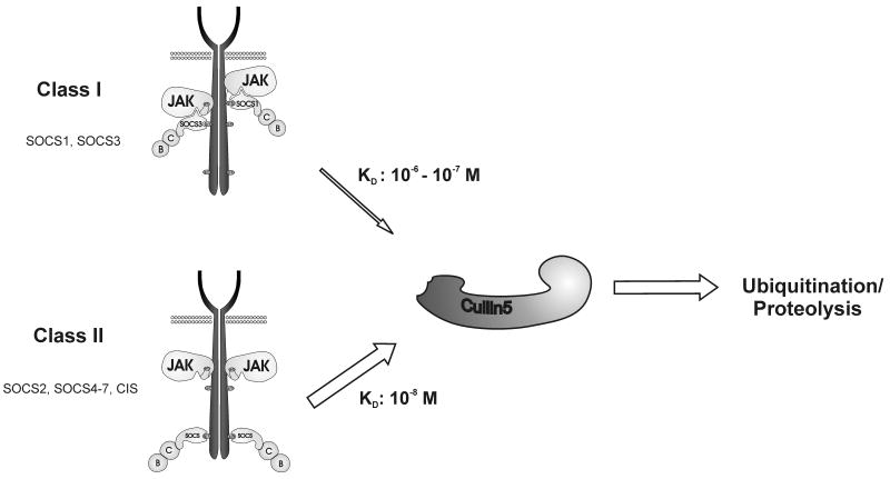
The SOCS (suppressors of cytokine signalling) family of proteins inhibits the cytokine-induced signalling cascade in part by promoting the ubiquitination of signalling intermediates that are then targeted for proteasomal degradation. This activity relies upon an interaction between the SOCS box domain, the adapter complex elonginBC and a member of the Cullin family, the scaffold protein of an E3 ubiquitin ligase. In this study, we dissected this interaction in vitro using purified components.We found that all eight SOCS proteins bound Cullin5 but required prior recruitment of elonginBC. Neither SOCS nor elonginBC bound Cullin5 when in isolation. Interestingly, the affinity of each SOCS-elonginBC complex for Cullin5 varied by 2 orders of magnitude across the SOCS family. Unexpectedly, the most potent suppressors of signalling, SOCS-1 and SOCS-3, bound most weakly to the E3 ligase scaffold, with affinities 100- and 10-fold lower, respectively, than the rest of the family. The remaining six SOCS proteins all bound Cullin5 with high affinity (K(d) of ~10 nM) due to a slower off-rate and hence a longer halflife of the complex. This difference in affinity may reflect a difference in mode of action as only SOCS-1 and SOCS-3 have been shown to suppress signalling using both SOCS box-dependent and SOCS box-independent mechanisms. This is not the case with the other six SOCS proteins, and our data imply the existence of two distinct subclasses of SOCS proteins with a high affinity for Cullin5, the E3 ligase scaffold, possibly reflecting complete dependence upon ubiquitination for suppression of cytokine signalling.


Figures

Figure 1
Figure 1. The SOCS box domain from all members of the SOCS family can bind elonginBC
(A) The SOCS box fragments were produced as GST fusion proteins and co-expressed with untagged elonginBC. Glutathione-Sepharose was used to pull down GST labelled proteins present in the cell lysate. As shown, the SOCS box domain from all eight members of the SOCS family could pull down elonginBC. Taking into account the different levels of SOCS expression there was no obvious difference in affinities among different members of the SOCS family. The asterisk indicates unavoidable protein degradation. (B) GST pulldowns such as those shown in (A) were digested with thrombin to remove the GST, then purified by gel filtration and concentrated to 10 mg/mL. In this case full-length SOCS3 was used as a positive control (lane S3), as the association of the SOCS3 box with elonginBC has already been described. The SOCS box of SOCS7 co-migrates with elonginC and so is not resolved by SDS-PAGE, however its presence was verified by mass-spectrometry (data not shown) and can be observed by NMR (Figure S1).
Figure 2
Figure 2. All members of the SOCS family, when in complex with elonginBC, bind cullin 5
(A) Ternary SOCS box/elonginBC complexes were incubated with glutathione Sepharose beads bound to a GST fusion of the N-terminal domain of cullin5. The SOCS box domains of all SOCS family members (S1-7, CIS), when bound to elonginBC, were shown to interact with cullin5. ElonginBC alone (BC) did not interact with cullin5. A fragment of the SOCS3 SOCS box, lacking the predicted cullin-binding motif (ΔC28) was used as a negative control (Δ). Asterisks mark GST-cullin5 degradation products, which are consistent across all experiments and were verified by mass spectrometry. Note that the SOCS7 box domain co-migrates with elonginC on SDS-PAGE. (B) Schematic view of our findings. Cullin5 binding requires a ternary SOCS/elonginB/elonginC complex. Neither elonginBC alone, nor a SOCS protein alone associate with the E3 ligase scaffold with measurable affinity.
Figure 3
Figure 3. The affinity for Cullin5 describes two sub-classes of SOCS protein
ITC analysis of the SOCS box/elonginBC-cullin5 interaction was used to measure the affinity of each SOCS box for Cullin5. (A) Tabular view of the results showing those proteins that interacted relatively weakly with Cullin5 (Class I), those that interacted strongly (KD ∼ 10 nM, Class II) and controls that did not interact at all. The values listed are the mean +/- S.D from two independent experiments using individually prepared batches of protein. The data for SOCS3/BC and elonginBC alone are from Babon et al (asterisks). (B) ITC data. 80 μM GST-cullin5 (NTD) was titrated into 10 μM SOCS box/elonginBC complex. The titration curves all fitted well to a single-site model with ∼ 1:1 stoichiometry within experimental error.
Figure 4
Figure 4. Analyses of the SOCS/elonginBC interaction
(A) Sequence comparison of the SOCS box from mouse CIS, SOCS1-7 and the three Drosophila SOCS proteins with the Cullin5-binding motif (LPLP) and other conserved residues shown highlighted in grey. (B) Mutating SOCS-1 and SOCS-3 to generate the canonical Cullin5 binding motif (LPLP) restores the affinity of Cullin5 for SOCS1 but not SOCS3. ITC analysis of the interaction between SOCS1(LPLP) and SOCS3 (LPLP) mutants with Cullin5. Because of a decreased enthalpy of interaction for both mutants the signal to noise ratio was low at 25°C (SOCS1(LPLP)=1.5 kcal/mol, Figure S2). The SOCS3(LPLP) interaction was too low to be measured, data not shown) so the reactions were repeated at 5°C where the interaction is endothermic in order to yield more accurate results. SOCS3 wild-type and mutant are shown in the lower two panels performed at 5°C. The titration curves all fitted well to a single-site model with ∼ 1:1 stoichiometry within experimental error. (C) Modelling of the SOCS2 box/elonginBC (PDB 2C9W) interaction with Cullin5 (right) using the Fbox-Skp2-Cullin1 structure (left) as a template (PDB 1LDK) is shown. The surface of Cullin is shown (grey) with hydrophobic residues in yellow and the ligands are represented as ribbon structures.
Figure 5
Figure 5. Surface plasmon resonance (Biacore) analyses of the SOCS/elonginBC-Cullin5 interaction
Sensorgrams of SOCS/elonginBC complexes binding to GST-Cullin5. Anti-GST antibody was covalently attached to a Biacore chip and used to capture GST-Cullin5 (NTD). Ternary SOCS/elonginBC complexes were then passed over the chip at 1, 0.5, 0.25, 0.125, 0.061 μM concentration (upper to lower curves in each panel). The curves were used to perform a global fit of the association and dissociation rates and the dissociation constant, the values are given below. All interactions could be fitted using a simple 1:1 Langmuir model with the exception of the SOCS1/elonginBC-Cullin5 interaction (top left) which appeared to have multiple binding components and was hence excluded from quantitation. All sensorgrams are plotted on the same scale for ease of comparison.
Figure 6
Figure 6. Schematic view of the division in the SOCS family based on mechanism and Cullin5 binding affinity
SOCS-1 and SOCS-3 define a separate class of SOCS proteins (termed Class I) based on their relatively low affinity for Cullin5 and the fact that they can interfere with signalling by associating with and inhibiting JAK. These two SOCS proteins are the most active at suppressing signalling and appear to be the most important regulators of the cytokine response to infection and inflammation. They are at least partially active in the absence of the SOCS box domain. The remaining SOCS proteins (class II) all bind tightly to Cullin5 and we suggest that the SOCS box is indispensable for their activity. Based on their higher affinity for the E3 ligase scaffold, class II SOCS proteins will be able to out-compete class I for cellular ubiquitination machinery. Both Classes of SOCS protein have to bind to elonginBC before the E3 ligase interaction will take place.

References

    1. Leonard WJ, O'Shea JJ. Jaks and STATs: biological implications. Annu Rev Immunol. 1998;16:293–322. - PubMed
    1. Starr R, Willson TA, Viney EM, Murray LJ, Rayner JR, Jenkins BJ, Gonda TJ, Alexander WS, Metcalf D, Nicola NA, Hilton DJ. A family of cytokine-inducible inhibitors of signalling. Nature. 1997;387:917–21. - PubMed
    1. Endo TA, Masuhara M, Yokouchi M, Suzuki R, Sakamoto H, Mitsui K, Matsumoto A, Tanimura S, Ohtsubo M, Misawa H, Miyazaki T, Leonor N, Taniguchi T, Fujita T, Kanakura Y, Komiya S, Yoshimura A. A new protein containing an SH2 domain that inhibits JAK kinases. Nature. 1997;387:921–4. - PubMed
    1. Naka T, Narazaki M, Hirata M, Matsumoto T, Minamoto S, Aono A, Nishimoto N, Kajita T, Taga T, Yoshizaki K, Akira S, Kishimoto T. Structure and function of a new STAT-induced STAT inhibitor. Nature. 1997;387:924–9. - PubMed
    1. Hilton DJ, Richardson RT, Alexander WS, Viney EM, Willson TA, Sprigg NS, Starr R, Nicholson SE, Metcalf D, Nicola NA. Twenty proteins containing a C-terminal SOCS box form five structural classes. Proc Natl Acad Sci U S A. 1998;95:114–9. - PMC - PubMed

Publication types

MeSH terms

Substances

LinkOut - more resources