. 2009 May;34(2):267-78.
doi: 10.1016/j.nbd.2009.01.013.

Anti-PrP Mab 6D11 suppresses PrP(Sc) replication in prion infected myeloid precursor line FDC-P1/22L and in the lymphoreticular system in vivo

Affiliations

Anti-PrP Mab 6D11 suppresses PrP(Sc) replication in prion infected myeloid precursor line FDC-P1/22L and in the lymphoreticular system in vivo

Martin J Sadowski et al. Neurobiol Dis. 2009 May.

Abstract

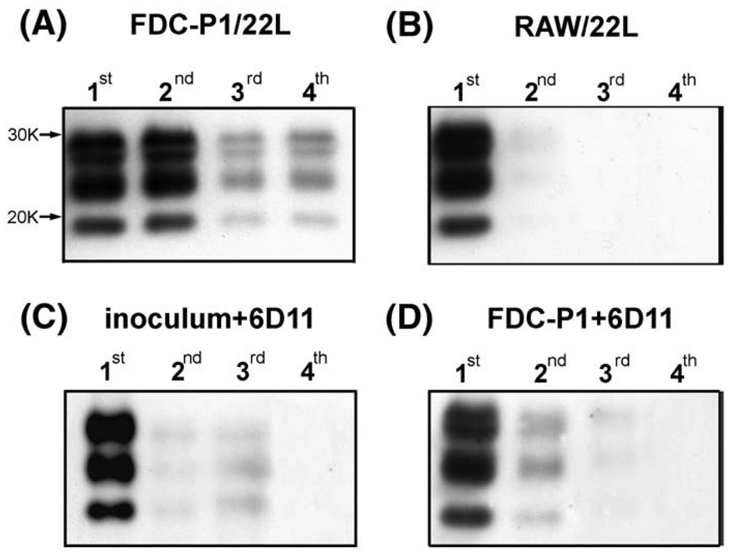
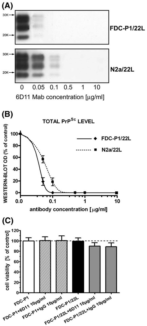
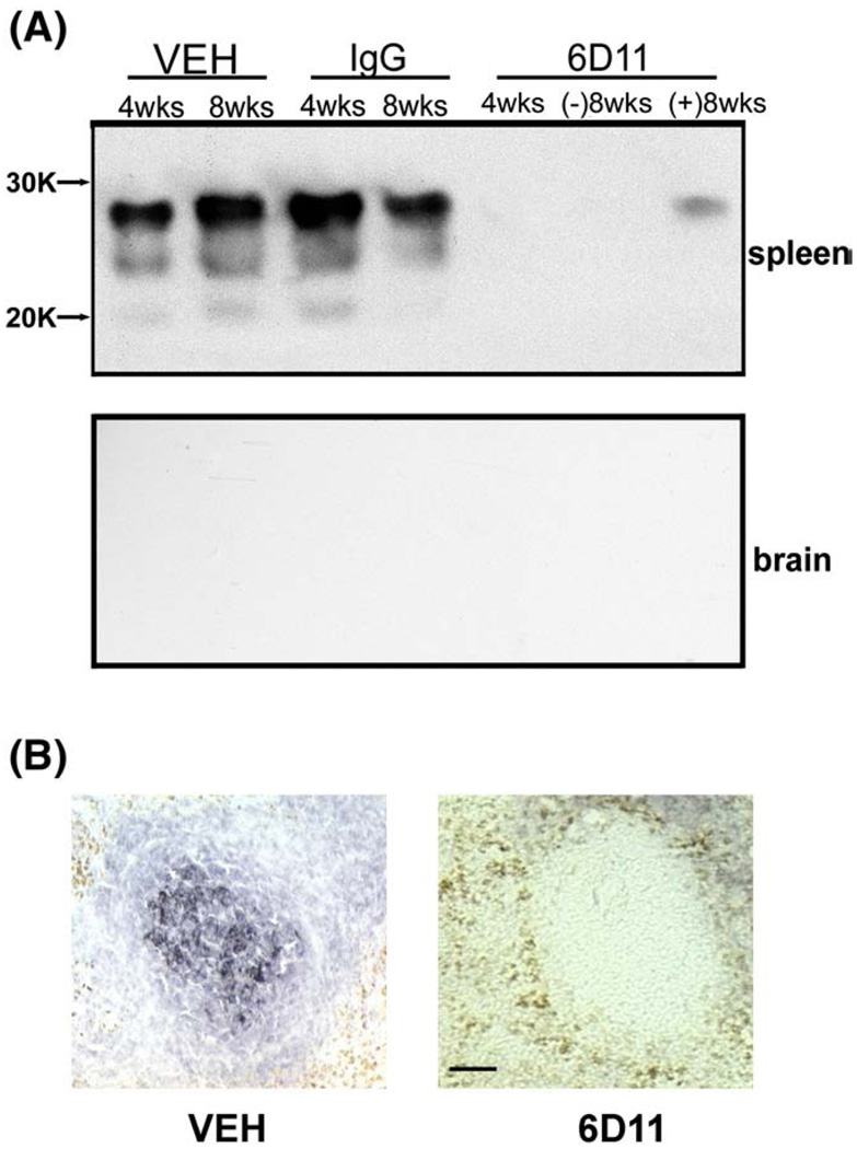
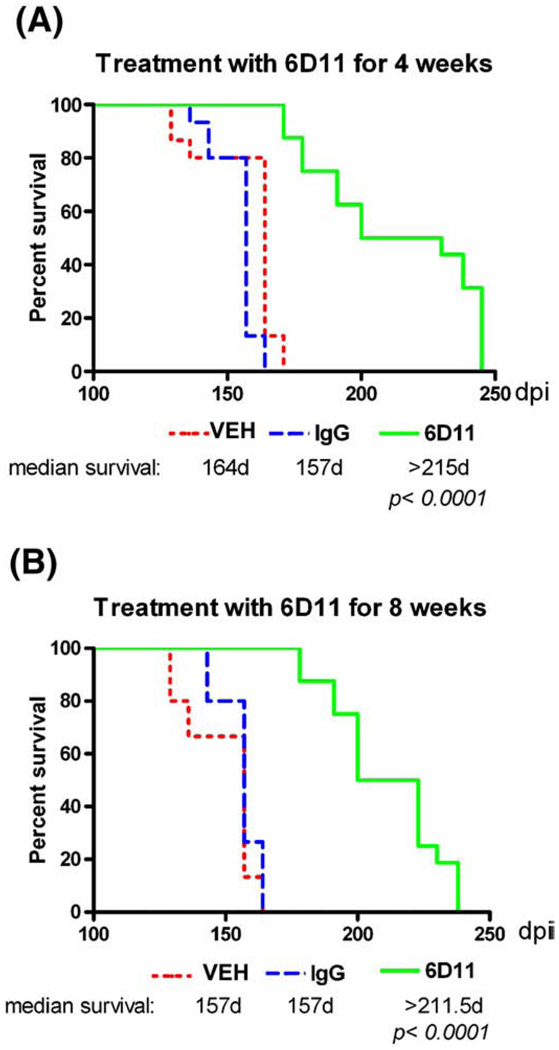
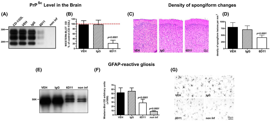
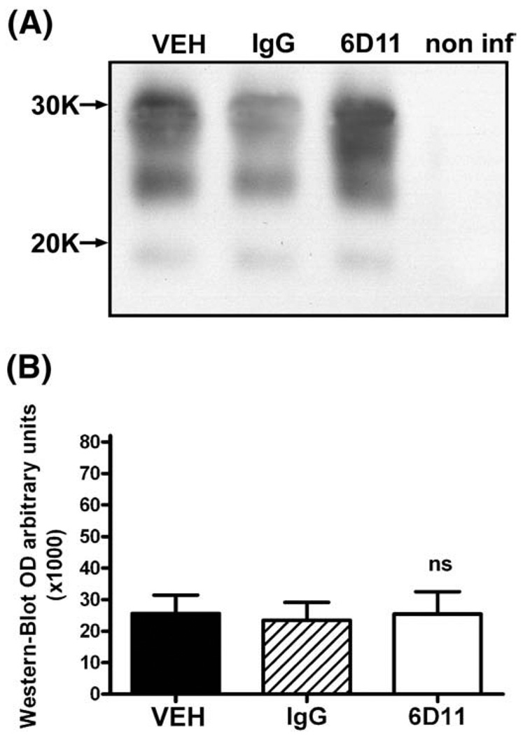
The pathogenesis of prion diseases is related to conformational transformation of cellular prion protein (PrP(C)) into a toxic, infectious, and self-replicating conformer termed PrP(Sc). Following extracerebral inoculation, the replication of PrP(Sc) is confined for months to years to the lymporeticular system (LRS) before the secondary CNS involvement results in occurrence of neurological symptoms. Therefore, replication of PrP(Sc), in the early stage of infection can be targeted by therapeutic approaches, which like passive immunization have limited blood-brain-barrier penetration. In this study, we show that 6D11 anti-PrP monoclonal antibody (Mab) prevents infection on a FDC-P1 myeloid precursor cell line stably infected with 22L mouse adapted scrapie strain. Passive immunization of extracerebrally infected CD-1 mice with Mab 6D11 resulted in effective suppression of PrP(Sc) replication in the LRS. Although, a rebound of PrP(Sc) presence occurred when the Mab 6D11 treatment was stopped, passively immunized mice showed a prolongation of the incubation period by 36.9% (pb0.0001) and a significant decrease in CNS pathology compared to control groups receiving vehicle or murine IgG. Our results indicate that antibody-based therapeutic strategies can be used, even on a short-term basis, to delay or prevent disease in subjects accidentally exposed to prions.


Figures

Fig. 1
Fig. 1
Characteristics of Mab 6D11. Mab 6D11 recognizes both human and murine PrPSc. (A) Shownis a comparison between the reactivity of 3F4 and 6D11 Mabs. Corresponding lanes represent PK digested brain homogenate from a: 1) GSS syndrome patient, 2) control human, 3) sporadic CJD, 4) AD patient, 5) CD-1 mouse infected with 139A mouse adapted scrapie strain, and 6) control mouse. (B) Comparison of homologous epitopes between sequences of murine and human PrP. 6D11 recognizes residues 97–100 of murine PrP (QWNK) which is homologous to sequence 98–101 of the human PrP. 3F4 recognizes residues 109–112 of human PrP (MKHM) but not homologous sequence of murine PrP (LKHV). The numbering of murine residues is shifted by one because murine PrP sequence lacks glycine in position 55. (C) Shown is a comparison of between binding affinities of 6D11 and 3F4 to human and murine recombinant PrP. Abbreviations: GSS—Gerstmann–Sträussler–Scheinker syndrome, ctrl hum—control human, sCJD—sporadic Creutzfeldt–Jakob disease, AD— Alzheimer's disease, CD—1/139A-CD-1 mouse infected with 139A mouse adapted scrapie strain, and ctrl-CD-1—control CD-1 mouse.
Fig. 2
Fig. 2
FDCs-P1 expresses a significant amount of PrPC. (A) Shown is the morphology and PrP expression in FDC-P1 and FDC-P1/22L cells. PrP expression in N2a murine neuroblastoma cells is shown for comparison. Cells were stained with 6D11 anti-PrP Mab (green) and counterstained with DAPI (blue). (B) Shown are confocal microscopy images (0.2 µm layer thickness) of FDC-P1 and FDC-P1/22L immunostained with 6D11 anti-PrP Mab. Anti-PrP immunoreactivity is primarily localized to the plasma membrane. No significant differences in staining intensity between non-infected and infected FDC-P1 cells were observed. (C) Shown is a comparison of the PrP content in cell lysates of N2a, FDC-P1 and FDC-P1/22L cells. Lane 1 represents N2a cells, lanes 2, 4, 6 FDC-P1 cells, and lanes 3, 5, 7 FDC-P1/22L cells. 20 µg of total protein was loaded in lanes 1, 4, 5; 30 µg in lanes 2 and 3 and 40 µg in lanes 6 and 7. Lysates from FDC-P1/22L cells were not PK digested. FDC-P1 cells express all three PrP isoforms with the diglycosylated form being the most abundant. The intensity of staining of 30 µg FDC-P1 and FDC-P1/22L samples (lanes 2 and 3) was comparable to that of 20 µg sample of N2a cell lysates (lane 1). No significant differences in the amount of total PrP between FDC-P1 and FDC-P1/22L lines were observed.
Fig. 3
Fig. 3
FDC-P1 cells are susceptible to infection with 22L strain in vitro and the infection can be prevented using 6D11 Mab. (A) Shown is a Western-blot of PK resistant PrPSc in passages 1–4 of FDC-P1 cells following infection with 22L strain. The presence of PK resistant material in passages 3 and 4 indicates the ability of FDC-P1 cells to sustain reproduction of PrPSc in vitro. (B) Shown are RAW cells (a macrophage line) that were exposed to PrPSc as a control experiment. These cells are able to phagocytize the inoculum but are unable to sustain de novo PrPSc replication, therefore PrPSc can be detected only in the first and partially in the second passage. (C) and (D) demonstrate the ability of Mab 6D11 to prevent infection of FDC-P1 line. In (C) the 22L inoculum was incubated with Mab 6D11 prior to infecting FDC-P1 cells, whereas in (D) FDC-P1 cells were incubated with Mab 6D11 prior to adding the 22L inoculum. Both types of intervention prevented effective infection since no PrPSc is detectable beyond passage 3.
Fig. 4
Fig. 4
Mab 6D11 abrogates the presence of PrPSc from N2a/22L and FDC-P1/22L infected cell lines without toxicity. (A) Shown are Western-blots of PK treated cell lysates from N2a/22L cells treated for 96 h with different concentrations of Mab. (B) Shown are densitometric measurements of PrPSc bands detected in the Western-blots and fitted in the sigmoidal dose–response curve. (C) Shown is MTTcytotoxicity assay on infected FDC-P1/22L cells. Neither infection of FDC-P1 line with 22L strain nor treatment of FDC-P1 or FDC-P1/22L lines with 6D11 or murine IgG produced significant decrease in cell viability as assessed by standard MTT cell viability assay. Values in (B) and (C) are given as a mean ± standard deviation from at least three independent experiments.
Fig. 5
Fig. 5
Systemic administration of 6D11 Mab inhibits replication of PrPSc in the LRS. (A) Shown is a highly sensitive immunoblotting assay used for detection of PrPSc in the spleen and the brain in sentinel mice sacrificed immediately after cessation of treatment. A substantial amount of PrPSc was detected in spleens of animals treated with vehicle or IgG that were sacrificed four or eight weeks after inoculation. No animal that received Mab 6D11 and was sacrificed four weeks after the inoculation was positive. Three out of five 6D11 treated animals were negative eight weeks after the inoculation ((−)8 wks), and the remaining two showed a faint signal ((+)8 wks). No animals showed the presence of PrPSc in the CNS eight weeks after the inoculation indicating that although PrPSc reaches a high concentration in the LRS there is no CNS involvement at that stage of infection. (B) Anti-PrP immunohistochemistry. Shown is accumulation of PrPSc in the germinal zone of the spleen in vehicle treated mice eight weeks after the inoculation. No PrPSc signal could be detected in Mab 6D11 treated mice at the matched time after inoculation. Scale bar 50 µm. Abbreviation: VEH—vehicle.
Fig. 6
Fig. 6
Short-term passive immunization delays the onset of neurological symptoms. Shown is a Kaplan–Meier analysis of the incubation time in CD-1 mice that were intraperitoneallyinfected with 22L strain and received treatment with 6D11 for (A) four or (B) eight weeks. A significant delay in onset of symptoms compared to the vehicle and IgG treated groups was observed in both experiments (p < 0.0001, log–rank test). Abbreviation: VEH—vehicle.
Fig. 7
Fig. 7
Short-term passive immunization has aneffecton CNS pathology. (A) and (B) show a significant reduction in PrPSc level in the CNS by semi-quantitative Western-blot. Values in (B) are expressed as a percentage of PrPSc level in the brain of CD-1 mice infected with 22L strain, which received no injections. Differences between 6D11 treated mice and 22L infected control mice, mice treated with vehicle or IgG were statistically significant as indicated. (C) shows representative hematoxylin and eosin stained cortical sections where reduced spongiform changes are evident in the 6D11 treated mice. (D) shows the reduction in the numerical density of spongiform lesions in the brain cortex in 6D11 treated mice. Differences between 6D11 treated mice, and mice treated with vehicle or IgG were statistically significant as indicated. Differences between mice treated with vehicle and IgG were not statistically significant. (E) and (F) show a significant reduction in GFAP level by semi-quantitative Western-blot. Differences between 6D11 treated mice, and mice treated with vehicle or IgG were statistically significant as indicated. Differences between GFAP level in non-infected mice and all other treatment groups (including 6D11) were significant as indicated. Differences in the GFAP level were associated with less activation of astrocytes as seen in (G) in representative GFAP immunohistochemically stained sections. Values in graphs (B), (D), and (F) are displayed as the mean ± standard deviation. Shown p values are for Tukey-HSD post-hoc test. Abbreviations: CD-1/22L—CD-1 mouse infected with 22L strain, non-inf—non infected, GFAP—glial fibrillary acidic protein, and VEH—vehicle.
Fig. 8
Fig. 8
The levels of PrPSc in the spleen did not differ significantly in control and 6D11 treated infected animals when they were killed after displaying neurological symptoms for three weeks in a row. Shown are (A) a Western-blot and (B) results of densitometric analysis of the PrPSc load in the spleen. No significant differences between 6D11 treated and control infected groups were detected. In (A) a non-infected spleen control is included to document the complete digestion of PrPC by PK treatment. Abbreviations: VEH—vehicle, non inf—non-infected, and PK—proteinase K.

Similar articles

Cited by

References

    1. Aucouturier P, Frangione B, Wisniewski T. Prion diseases and the immune system. Ann. Med. Interne. 1999;150:75–78. - PubMed
    1. Aucouturier P, Geissmann F, Damotte D, Saborio GP, Meeker HC, Kascsak R, Kascsak R, Carp RI, Wisniewski T. Infected dendritic cells are sufficient for prion transmission to the CNS in mouse scrapie. J. Clin. Invest. 2001;108:703–708. - PMC - PubMed
    1. Beekes M, McBride PA. Early accumulation of pathological PrP in the enteric nervous system and gut-associated lymphoid tissue of hamsters orally infected with scrapie. Neurosci. Lett. 2000;278:181–184. - PubMed
    1. Beekes M, McBride PA, Baldauf E. Cerebral targeting indicates vagal spread of infection in hamsters fed with scrapie. J. Gen. Virol. 1998;3:601–607. - PubMed
    1. Bourette RP, Myles GM, Carlberg K, Chen AR, Rohrschneider LR. Uncoupling of the proliferation and differentiation signals mediated by the murine macrophage colony-stimulating factor receptor expressed in myeloid FDC-P1 cells. Cell Growth Differ. 1995;6:631–645. - PubMed

Publication types

MeSH terms

LinkOut - more resources