. 2009 May;34(2):279-90.
doi: 10.1016/j.nbd.2009.01.016.

A highly reproducible rotenone model of Parkinson's disease

Affiliations

A highly reproducible rotenone model of Parkinson's disease

Jason R Cannon et al. Neurobiol Dis. 2009 May.

Abstract

The systemic rotenone model of Parkinson's disease (PD) accurately replicates many aspects of the pathology of human PD and has provided insights into the pathogenesis of PD. The major limitation of the rotenone model has been its variability, both in terms of the percentage of animals that develop a clear-cut nigrostriatal lesion and the extent of that lesion. The goal here was to develop an improved and highly reproducible rotenone model of PD. In these studies, male Lewis rats in three age groups (3, 7 or 12-14 months) were administered rotenone (2.75 or 3.0 mg/kg/day) in a specialized vehicle by daily intraperitoneal injection. All rotenone-treated animals developed bradykinesia, postural instability, and/or rigidity, which were reversed by apomorphine, consistent with a lesion of the nigrostriatal dopamine system. Animals were sacrificed when the PD phenotype became debilitating. Rotenone treatment caused a 45% loss of tyrosine hydroxylase-positive substantia nigra neurons and a commensurate loss of striatal dopamine. Additionally, in rotenone-treated animals, alpha-synuclein and poly-ubiquitin positive aggregates were observed in dopamine neurons of the substantia nigra. In summary, this version of the rotenone model is highly reproducible and may provide an excellent tool to test new neuroprotective strategies.


Figures

Figure 1
Figure 1
Daily intraperitoneal rotenone elicits moderate weight loss and a parkinsonian phenotype. A-C.,Percent change in mass from day 0 in (A) young-adult rats (∼ 3 months old; control, n = 13; 2.75 mg/kg/day, n = 7; 3.0, n = 19), (B) adult rats (∼7 months; control, n = 5; 2.75 mg/kg/day, n = 7; 3.0, n = 6) and (C) middle-aged rats (12-14 months, control, n = 5; 2.75 mg/kg/day, n = 5; 3.0, n = 6) treated with rotenone at 2.75 mg/kg/day (empty circles), 3.0 mg/kg/day (filled circles) and control rats (filled squares). D-F, Survival curves (development of debilitating PD phenotype characterized by severe bradykinesia, rigidity, and postural instability) of young-adult (D), adult (E) and middle-aged (F) rats treated with rotenone (2.75 or 3.0 mg/kg/day) and control rats. Animals were euthanized when severe parkinsonian symptoms developed. Mortality occurring immediately after the injection (in the absence of parkinsonian symptoms and typically <10%) was excluded from analysis. Note that the percent mass change is relatively similar for all age groups at specific time-points and dosages. Young-adult animals exhibit the most variability in temporal development of parkinsonian phenotype. Adult animals exhibit a dose-dependent response to rotenone (* = p<0.05, Log rank test for trend). Middle-aged animals also appear to be the most sensitive to rotenone.
Figure 2
Figure 2
Neurobehavioral assessment of rotenone-treated rats. Daily intraperitoneal rotenone (3.0 mg/kg/day) in young-adult rats elicits progressive motor deficits that are responsive to a dopamine agonist. A, Postural instability test at pretest, and at 3, 6, and 9 days of rotenone treatment (empty circles represent control, n = 7; filled circles represent rotenone-treated, n = 13). ***p<0.001, Student's t-test. B, Postural instability test before and after apomorphine (1 mg/kg) challenge. ***p<0.001, paired Student's t-test (pre-apomorphine versus post-apomorphine). C, Rearing behavior (# of rears per 5 min). *p<0.05, **p<0.01, Student's t-test. D, Rearing before and after apomorphine challenge. **p<0.01, ***p<0.001, paired Student's t-test (pre-apomorphine versus post-apomorphine).
Figure 3
Figure 3
Intraperitoneal rotenone elicits dopamine terminal loss in the dorsolateral striatum. Representative images (from 3 treated animals in each group) of striatal tyrosine hydroxylase immunohistochemistry in young-adult (A-D), adult (E-H), and middle-aged (I-L) rats after daily intraperitoneal rotenone (3.0 mg/kg/day, A-C, E-G, I-K) or vehicle (D,H,L). Note that the lesions are strikingly similar in magnitude and localization across animals, particularly in the middle-aged animals. Bar = 500 μm.
Figure 4
Figure 4
Rotenone elicits striatal dopamine terminal hypertrophy. Representative high magnification images of dorsolateral striatal tyrosine hydroxylase immunohistochemistry in vehicle (A-C) or rotenone [2.75 mg/kg/day (D-F), 3.0 mg/kg/day (G-I)] treated middle-aged animals. Arrows indicate hypertrophied and likely dysfunctional dopaminergic terminals. Hypertrophied terminals were present in all treated animals, both near the edge of the lesion in animals treated at 3.0 mg/kg and also in animals treated at 2.75 mg/kg rotenone, without an overt nigrostriatal lesion. Bar = 10 μm.
Figure 5
Figure 5
Sparing of striatal neurons after intraperitoneal rotenone. Images show Nissl staining and tyrosine immunohistochemistry in the dorsolateral striatum of control (A-C) and rotenone-treated middle-aged rats (3.0 mg/kg/day; D-F). Rotenone-treated animals exhibit a striking loss of striatal tyrosine hydroxylase immunoreactivity, while striatal cell bodies are spared, as evidenced by persistence of Nissl stained striatal neurons. Bar = 50 μm. High magnification inserts (Ci and Fi) show no alterations in striatal cell morphology or evidence of inflammatory cell infiltration in rotenone treated (Fi) compared to control (Ci). Bar = 25 μm.
Figure 6
Figure 6
Striatal DARPP-32 staining. Striatal staining for tyrosine hydroxylase (TH) and DARPP-32. Low magnification images show that there is loss of TH+ terminals in rotenone treated animals with no overt loss of DARPP-32+ striatal cell bodies. High magnification images show that DARPP-32+ neurons do not exhibit morphological alterations in rotenone treated animals.
Figure 7
Figure 7
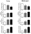
Intraperitoneal rotenone causes striatal dopamine depletion in young and middle-aged rats. Dopamine (DA), 3,4-dihydroxyphenylacetic acid (DOPAC), homovanillic acid (HVA) (ng/mg protein), and dopamine turnover [(DOPAC+HVA)/DA] in rotenone- (2.75, 3.0 mg/kg/day) and vehicle-treated rats. n = 5-7 per group. Note that alterations in catecholamine levels are similar in both age groups. * p<0.025, ** p<0.01, *** p<0.001, compared to control. Student's t-test with Bonferroni correction for multiple comparisons.
Figure 8
Figure 8
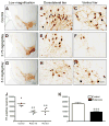
Intraperitoneal rotenone results in the loss of nigral tyrosine hydroxylase- (TH) positive neurons in middle-aged rats. Tyrosine hydroxylase immunohistochemistry (A-I) in control (A-C), and rotenone-treated rats [2.75 mg/kg/day (D-F) or 3.0 mg/kg/day (G-I)]. Low magnification images show overall cell loss and pruning of processes (A,D,G) (bar = 500 μm), medium magnification images in dorsolateral and ventral regions of nigra show cell loss, morphological changes, and loss of processes in these particularly vulnerable areas (bar = 50 μm). Unbiased stereological cell counts (J,K) show quantitative measurement of cell loss. ANOVA with Tukey's post hoc test indicated that both rotenone doses caused significant loss of dopamine neurons. Because no differences were found between animals treated at 2.75 or 3.0 mg/kg/day (J), these groups were merged and subjected to a student's t-test (K). **p<0.01; ***p<0.001 compared to control.
Figure 9
Figure 9
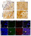
Intraperitoneal rotenone-induces α-synuclein and poly-ubiquitin accumulation and aggregation. Nigral α-synuclein immunohistochemistry in proteinase K pretreated (A,C) and formic acid pretreated tissue sections (B,D) from control (A,B) or rotenone-treated (3.0 mg/kg/day, C,D) animals (bars = 25 μm). E-L, Confocal microscopy of triple-labeled immunofluorescence for α-synuclein (α-syn; E,I), poly-ubiquitin (poly-ubiq; F,J), and pSer19-tyrosine hydroxylase (TH; G,K) and merged image (H,L) in a control (E-H) and rotenone-treated rat (I-L). E-L (bar = 10 μm). In the rotenone-treated rat, a dopamine neuron can be seen to contain multiple, large, round inclusions that stain for both α-synuclein and poly-ubiquitin.

Similar articles

Cited by

References

    1. Alam M, Schmidt WJ. Rotenone destroys dopaminergic neurons and induces parkinsonian symptoms in rats. Behav Brain Res. 2002;136:317–24. - PubMed
    1. Alam M, Schmidt WJ. L-DOPA reverses the hypokinetic behaviour and rigidity in rotenone-treated rats. Behav Brain Res. 2004;153:439–46. - PubMed
    1. Betarbet R, Canet-Aviles RM, Sherer TB, Mastroberardino PG, McLendon C, Kim JH, Lund S, Na HM, Taylor G, Bence NF, Kopito R, Seo BB, Yagi T, Yagi A, Klinefelter G, Cookson MR, Greenamyre JT. Intersecting pathways to neurodegeneration in Parkinson's disease: effects of the pesticide rotenone on DJ-1, alpha-synuclein, and the ubiquitin-proteasome system. Neurobiol Dis. 2006;22:404–20. - PubMed
    1. Betarbet R, Sherer TB, MacKenzie G, Garcia-Osuna M, Panov AV, Greenamyre JT. Chronic systemic pesticide exposure reproduces features of Parkinson's disease. Nat Neurosci. 2000;3:1301–6. - PubMed
    1. Biehlmaier O, Alam M, Schmidt WJ. A rat model of Parkinsonism shows depletion of dopamine in the retina. Neurochem Int. 2007;50:189–95. - PubMed

Publication types

MeSH terms

LinkOut - more resources