. 2009;11(2):R20.
doi: 10.1186/bcr2245. Epub 2009 Apr 23.

Pregnancy in the mature adult mouse does not alter the proportion of mammary epithelial stem/progenitor cells

Affiliations

Pregnancy in the mature adult mouse does not alter the proportion of mammary epithelial stem/progenitor cells

Kara L Britt et al. Breast Cancer Res. 2009.

Abstract

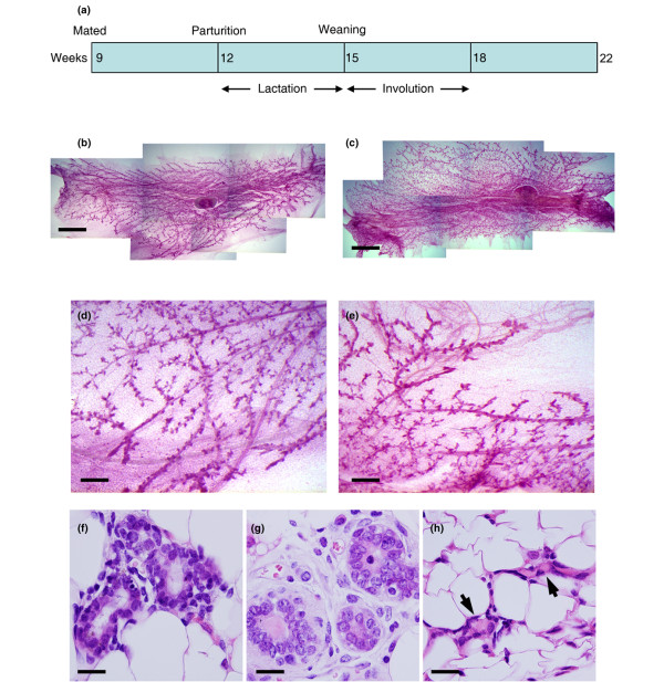
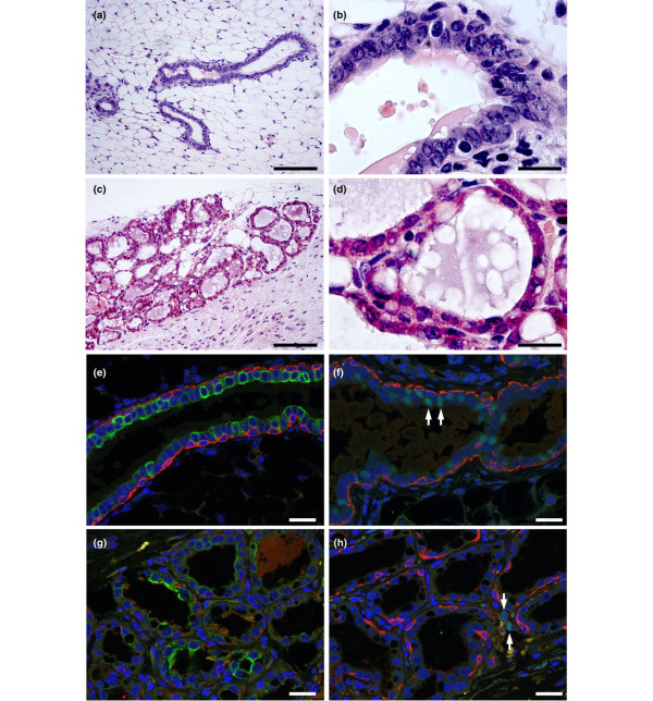
Introduction: In humans, an early full-term pregnancy reduces lifetime breast cancer risk by up to 50% whereas a later pregnancy (>35 years old) can increase lifetime risk. Several mechanisms have been suggested, including changes in levels of circulating hormones, changes in the way the breast responds to these hormones, changes in gene expression programmes which may alter susceptibility to transformation and changes to mammary stem cell numbers or behaviour. Previous studies have shown that the mammary tissue isolated from both virgin and parous mice has the ability to repopulate a cleared mammary fat pad in transplant experiments. Limited dilution transplant assays have demonstrated that early pregnancy (at 5 weeks of age) reduces stem/progenitor cell numbers in the mouse mammary epithelium by twofold. However, the effects on stem/progenitor cell numbers in the mammary epithelium of a pregnancy in older animals have not yet been tested.

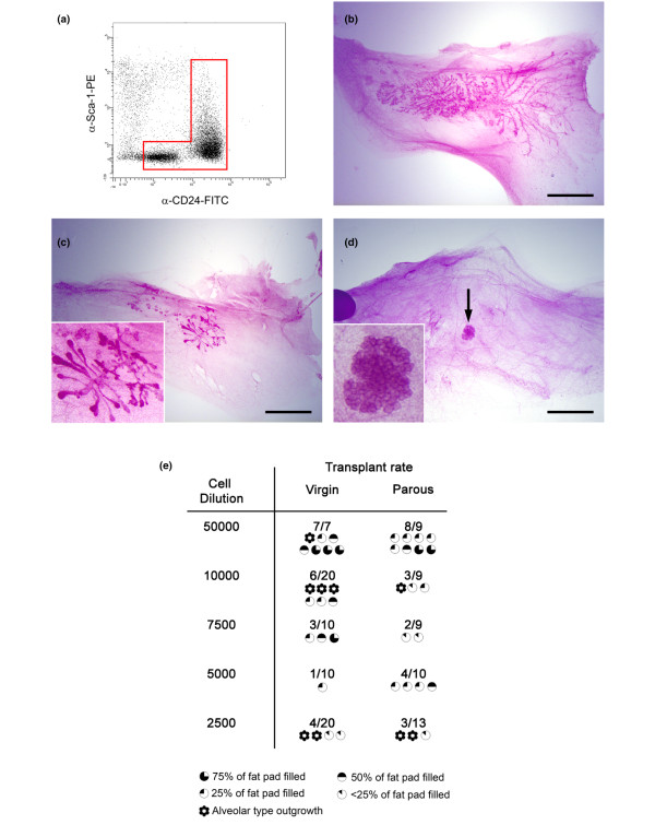
Methods: Mice were put through a full-term pregnancy at 9 weeks of age, when the mammary epithelium is mature. The total mammary epithelium was purified from parous 7-week post-lactation and age-matched virgin mice and analysed by flow cytometry and limiting dilution cleared fat pad transplants.

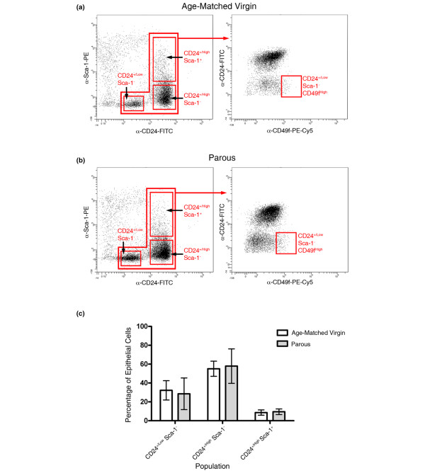
Results: There were no significant differences in the proportions of different mammary epithelial cell populations or numbers of CD24(+/Low) Sca-1- CD49f(High) cells (stem cell enriched basal mammary epithelial compartment). There was no significant difference in stem/progenitor cell frequency based on limiting dilution transplants between the parous and age-matched virgin epithelium.

Conclusions: Although differences between parous and virgin mammary epithelium at later time points post lactation or following multiple pregnancies cannot be ruled out, there are no differences in stem/progenitor cell numbers between mammary epithelium isolated from parous animals which were mated at 9 weeks old and virgin animals. However, a recent report has suggested that animals that were mated at 5 weeks old have a twofold reduction in stem/progenitor cell numbers. This is of interest given the association between early, but not late, pregnancy and breast cancer risk reduction in humans. However, a mechanistic connection between stem cell numbers and breast cancer risk remains to be established.


Figures

Figure 1
Figure 1
Seven-week post-lactation mouse mammary epithelium is fully regenerated and similar to age-matched virgin tissue. (a) Outline of experimental design. Mice were mated at 9 weeks of age, went through a normal pregnancy and nursed pups for 3 weeks. Involution occurred over an approximately 3-week period after pups were removed. After a further 4 weeks, when the animals were in total 22 weeks old, tissue was harvested. (b), (d) Carmine-stained whole mount of age-matched virgin (AMV) fourth mammary fat pad. (c), (e) Carmine-stained whole mount of parous fourth mammary fat pad. (b), (c) Bar = 3 mm. (d), (e) Bar = 750 μm. (f) H & E-stained section of AMV fourth mammary fat pad. Bar = 20 μm. (g) H & E-stained section of parous (7-week post-lactation) fourth mammary fat pad. Bar = 20 μm. (h) H & E-stained section of parous (3-week post-lactation) fourth mammary fat pad showing collapsed ducts (arrowheads). Bar = 20 μm.
Figure 2
Figure 2
CD24 and Sca-1 flow cytometry isolates similar populations in age-matched virgin and parous tissues. (a) Age-matched virgin (AMV) cells and (b) parous cells were stained with antibodies against CD24, Sca-1 and CD49f. The CD24 and Sca-1 plots (left panels) identified CD24+/Low Sca-1-, CD24+/High Sca-1- and CD24+/High Sca-1+ cells in both AMV and parous cell preparations. Analysis of these populations for CD49f expression (right panels) enabled identification of the CD24+/Low Sca-1- CD49fHigh population previously shown to be enriched for mammary stem cells [11,26,27]. See also Additional data file 1. (c) Relative abundance of CD24+/Low Sca-1-, CD24+/High Sca-1- and CD24+/High Sca-1+ cells as a percentage of the total epithelium in AMV and parous tissue.
Figure 3
Figure 3
Isolation of total epithelial cell populations from mammary epithelium. (a) Whole mount of mammary fat pad transplanted with primary cells transduced with green fluorescent protein (GFP) lentivirus. Arrow, area enlarged in inset. Bar = 2.5 mm. (b) Flow cytometric analysis of transplanted epithelium. The histogram indicates levels of GFP fluorescence in cells isolated from transplanted mammary fat pads. The CD24 Sca-1 staining pattern of the total cells (GFP- and GFP+) harvested from the fat pads is shown. The CD24 Sca-1 staining patterns of the GFP- and GFP+ cells are also shown separately. GFP+ cells, which must represent the progeny of transduced stem cells, are found only in the CD24+/Low Sca-1-, CD24+/High Sca-1- and CD24+/High Sca-1+ regions. Data are representative of four independent experiments.
Figure 4
Figure 4
Proportion of stem/progenitor cells in mammary epithelium unchanged in parous compared with age-matched virgin tissue. (a) Flow cytometry plot of cells isolated from age-matched virgin (AMV) tissue and stained with anti-CD24 and anti-Sca-1 antibodies showing a typical gate used to isolate total mammary epithelial cells for transplantation. This gate was based on the data obtained from the lentiviral transduction experiments, which demonstrated that the CD24+/Low Sca-1- plus the total CD24+/High regions defined the entire mammary epithelium. (b) to (d) Carmine stained whole mounts showing examples of (b) large ductal outgrowths, (c) small ductal outgrowths and (d) an alveolar structure generated by transplantation. Arrow, region enlarged in the inset. Bar = 4 mm. (e) Table of results of limiting dilution transplants indicating the number of outgrowths obtained and the number of fat pads transplanted for each cell dilution. The extent to which each outgrowth filled the host fat pad is indicated by the symbols below the numbers. Data are from seven independent cell isolation and transplant sessions.
Figure 5
Figure 5
Outgrowths of transplanted parous and age-matched virgin tissue. Outgrowths of transplanted parous and age-matched virgin tissue contain basal/myoepithelial cells, luminal oestrogen receptor-negative (ER-) cells and luminal oestrogen receptor-positive (ER+) cells. (a), (b) H & E-stained section of ductal-type transplant outgrowth. (c), (d) H & E-stained section of alveolar-type transplant outgrowth. (a), (c) Bar = 120 μm. (b), (d) Bar = 20 μm. (e) to (h) Sections of outgrowth stained by multiple immunofluorescence for cell-type specific markers. All nuclei are counterstained with DAPI (blue). Bar = 20 μm. (e) Ductal outgrowth stained for keratin 14 (red) and keratin 18 (green). (f) Ductal outgrowth stained for keratin 14 (red) and ER (green). Arrows, examples of ER+ luminal epithelial nuclei. (g) Alveolar outgrowth stained for keratin 14 (red) and keratin 18 (green). (h) Alveolar outgrowth stained for keratin 14 (red) and ER (green). Arrows indicate rare interstitial ER+ cells between alveolar structures.

References

    1. MacMahon B, Cole P, Lin TM, Lowe CR, Mirra AP, Ravnihar B, Salber EJ, Valaoras VG, Yuasa S. Age at first birth and breast cancer risk. Bull World Health Organ. 1970;43:209–221. - PMC - PubMed
    1. Britt K, Ashworth A, Smalley M. Pregnancy and the risk of breast cancer. Endocr Relat Cancer. 2007;14:907–933. doi: 10.1677/ERC-07-0137. - DOI - PubMed
    1. Albrektsen G, Heuch I, Hansen S, Kvale G. Breast cancer risk by age at birth, time since birth and time intervals between births: exploring interaction effects. Br J Cancer. 2005;92:167–175. doi: 10.1038/sj.bjc.6602302. - DOI - PMC - PubMed
    1. Schedin P. Pregnancy-associated breast cancer and metastasis. Nat Rev Cancer. 2006;6:281–291. doi: 10.1038/nrc1839. - DOI - PubMed
    1. Trichopoulos D, Hsieh CC, MacMahon B, Lin TM, Lowe CR, Mirra AP, Ravnihar B, Salber EJ, Valaoras VG, Yuasa S. Age at any birth and breast cancer risk. Int J Cancer. 1983;31:701–704. doi: 10.1002/ijc.2910310604. - DOI - PubMed

Publication types