

Synovial chondromatosis originating from the synovium of the anterior cruciate ligament: a case report
- PMID: 19386135
- PMCID: PMC2678102
- DOI: 10.1186/1758-2555-1-6
Synovial chondromatosis originating from the synovium of the anterior cruciate ligament: a case report
Abstract
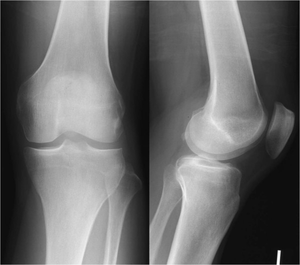
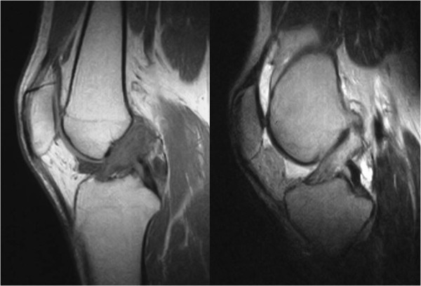
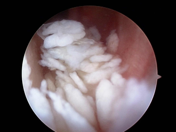
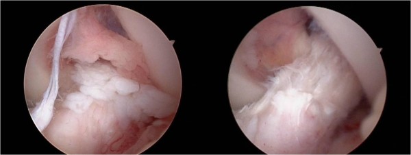
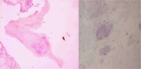
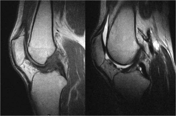
A case of synovial chondromatosis originating from the synovium of the anterior cruciate ligament (ACL) resulting in a mechanical block to knee extension is reported. A 36-year-old man complained of a restricted range of left-knee motion and pain when walking. Plain roentgenograms showed normal appearance, however, magnetic resonance imaging showed intensity changes in the ACL. Arthroscopically, numerous small free bodies were observed. Proliferation of synovium and cartilaginous tissues were identified around the ACL. There were no significant findings in the synovium except around the ACL. The synovium around the ACL was resected and free bodies were washed out. This is the first report of synovial chondromatosis originating from the synovium of the ACL.
Figures
References
-
- Jeffreys TE. Synovial Chondromatosis. J Bone Joint Surg Br. 1967;49:530–4. - PubMed
-
- Maurice H, Crone M, Watt I. Synovial Chondromatosis. J Bone Joint Surg Br. 1988;70:807–11. - PubMed
-
- Mubashir A, Bickerstaff DR. Synovial Osteochondromatosis of the Cruciate Ligament. Arthroscopy. 1998;14:627–629. - PubMed
-
- Kay PR, Freemont AJ, Davies DRA. The Etiology of multiple loose bodies. Snow Storm Knee. J Bone Joint Surg Br. 1989;71:501–504. - PubMed
LinkOut - more resources
Full Text Sources
