. 2009 Apr;16(4):539-50.
doi: 10.1016/j.devcel.2009.02.004.

Plk1-dependent and -independent roles of an ODF2 splice variant, hCenexin1, at the centrosome of somatic cells

Affiliations

Plk1-dependent and -independent roles of an ODF2 splice variant, hCenexin1, at the centrosome of somatic cells

Nak-Kyun Soung et al. Dev Cell. 2009 Apr.

Abstract

Outer dense fiber 2 (ODF2) was initially identified as a major component of the sperm tail cytoskeleton, and was later suggested to be localized to somatic centrosomes and required for the formation of primary cilia. Here we show that a splice variant of hODF2 called hCenexin1, but not hODF2 itself, efficiently localizes to somatic centrosomes via a variant-specific C-terminal extension and recruits Plk1 through a Cdc2-dependent phospho-S796 motif within the extension. This interaction and Plk1 activity were important for proper recruitment of pericentrin and gamma-tubulin, and, ultimately, for formation of normal bipolar spindles. Earlier in the cell cycle, hCenexin1, but again not hODF2, also contributed to centrosomal recruitment of ninein and primary cilia formation independent of Plk1 interaction. These findings provide a striking example of how a splice-generated C-terminal extension of a sperm tail-associating protein mediates unanticipated centrosomal events at distinct stages of the somatic cell cycle.


Figures
Fig. 1
Fig. 1
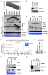
Plk1 binds to the C-terminal region of hCenexin1 during the late stages of the cell cycle. (A), HeLa cells were infected with adenovirus expressing either EGFP-hCenexin1 or EGFP-ODF2 and then subjected to confocal microscopy (Left). Arrows indicate centrioles. Because of a low background signals for EGFP-hCenexin1, a circle was drawn to indicate the position of nuclear membrane. Relative fluorescence intensities for centrosome-localized EGFP-hCenexin1 (n = 29 cells) and EGFP-ODF2 (n = 23 cells) signals over cytoplasmic background were quantified (Right). Bars (red), the averages of relative fluorescence intensities. (B), Total cellular lysates from mitotic HeLa cells were fractionated using a ZZ tag-affinity column. Proteins associated with hCenexin1 or hODF2 were eluted after TEV digestion, and then analyzed. (C), Total cellular lysates were prepared from HeLa cells infected with the indicated adenoviruses and treated with nocodazole for 16 h. Samples were subjected to PBD pull-downs as described in the Experimental procedures. After immunoblotting, the membrane was stained with Coomassie (CBB) to determine the levels of ligands precipitated. (D), Total lysates prepared from mitotic HeLa cells were subjected to PBD pull-downs with bead-bound wild-type GST-PBD (WT) or the GST-PBD(H538A K540M) mutant (AM). Asterisks, non-specific cross-reacting proteins. (E), HeLa cells released from double thymidine block into fresh medium were harvested for PBD pull-downs. Cells silenced for hCenexin1 (sh-781) or treated with nocodazole (Noc) were included for comparison. Asterisks, cross-reacting proteins. (F), Total cellular proteins were prepared from HeLa cells growing asynchronously (Asyn) or treated with nocodazole for 16 h (Noc). To reveal a slow-migrating hCenexin1 form, samples were long separated by 7.5% low-bis (96:1 acrylamide:bis-acrylamide) SDS-PAGE and immunoblotted (Left). Mitotic HeLa lysates (50 μg) were either treated with λ-phosphatase or left untreated and analyzed (Right). Asterisks, cross-reacting proteins.
Fig. 2
Fig. 2
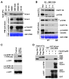
Phosphorylation of hCenexin1 at S796 is critical for the hCenexin1-Plk1 interaction. (A), HeLa cells expressing various EGFP-hCenexin1 forms were treated with nocodazole for 16 h, harvested, and then subjected to PBD pull-downs. Numbers indicate relative efficiencies of PBD-binding in comparison to the levels of input. Arrows, weakly detectable hCenexin1 proteins; Asterisk, EGFP-T7 stained with Coomassie. (B), Mitotic HeLa lysates expressing EGFP-T8 were incubated with wild-type GST-PBD (WT) or the GST-PBD(H538A K540M) mutant (AM), and then analyzed. (C), To determine in vivo phosphorylation site(s) on the C-terminal region of hCenexin1, EGFP-T7 immunoprecipitated from nocodazole-treated HeLa cells (inset gel) was subjected to mass spectrometry analysis as described in the Experimental procedures. The phosphorylation site was determined by the phosphorylated (red) fragment ions with (w/) or without (w/o) neutral loss of phosphate and unphosphorylated (blue) fragment ions. (D), Mitotic HeLa cells expressing various hCenexin1 forms were subjected to PBD pull-downs. To mark the positions of the endogenous and exogenous hCenexin1 proteins, samples expressing wild-type hCenexin1 (WT) or vector but depleted of endogenous hCenexin1 (sh-781) (lanes 6 and 7) were included. Due to a cloning variation at the N-terminal region, the exogenous hCenexin1 migrated about 5-kDa faster than the endogenous hCenexin1. Asterisks, non-specific cross-reacting proteins. (E), A pair of short N-terminal Cys-containing non-phospho- and phospho-S796 peptide was cross-linked to the beads and then incubated with mitotic HeLa lysates. The resulting peptide-associated cellular proteins were analyzed. (F), Mitotic HeLa lysates were preincubated with buffer (−), non-phospho-, or phospho-S796 peptide for 20 min, and then subjected to PBD pull-downs. Likely due to phosphorylation, migration of the PBD-bound hCenexin1 was often distinguishably slower than that of hCenexin1 in the input. Asterisk, a cross-reacting protein. (G), Mitotic HeLa cells expressing EGFP-Plk1 were preincubated with buffer (−) or the indicated peptide prior to immunoprecipitation.
Fig. 3
Fig. 3
Cdc2-dependent phosphorylation of hCenexin1 at S796 is sufficient to induce the hCenexin1-Plk1 interaction. (A), Mitotic HeLa cells treated with the indicated inhibitors for 30 min were harvested and subjected to PBD pull-downs. Asterisks, non-specific cross-reacting proteins. (B), HeLa cells stably expressing EGFP-T8(689–805) were arrested at the G1/S boundary by double thymidine treatment, released into fresh medium for 3 h, and then treated with etoposide for 7 h to re-arrest the cells in G2. BMI-1026 was added into the etoposide-containing medium 1 h or 2 h before harvest. Nocodazole-arrested cells (Noc) were included for comparison. To determine the level of phospho-independent binding, nocodazole-treated lysates from lane 1 were also pulled down with the PBD(H538A K540M) (AM) mutant. Numbers, the level of p-S796 EGFP-T8 over total EGFP-T8 relative to the 0 h sample. Asterisk, a cross-reacting protein. (C), HeLa cells expressing EGFP-T8 were infected with adenoviruses encoding Cdc2 and Cyclin B1, and then arrested at the G1/S boundary before harvest. Samples were immunoblotted with anti-p-S796 antibody preincubated with the indicated peptides or with anti-GFP antibody to determine the level of total EGFP-T8. (D), Bead-bound control GST or GST-hCenexin1(647–805) were reacted with purified Cdc2/Cyclin B1 complex in the presence or absence of ATP. After washing, the resulting ligands were incubated with a 1:1:1 mixture of transfected lysates for Flag-Plk1, Flag-Plk2, and Flag-Plk3, and then the ligand-associating proteins were analyzed. Asterisk, a cross-reacting protein.
Fig. 4
Fig. 4
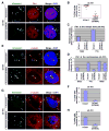
Overexpressed hCenexin1, but not hCenexin1(S796A), forms hCenexin1-Plk1 assemblies, recruits γ-tubulin, and generates ectopic microtubule organizing centers. (A–F), HeLa cells silenced for hCenexin1 (sh-781) were arrested by double thymidine block and released for 10 h (DT 10 h) to enrich the G2 population. Upon releasing from the block, cells were infected with adenovirus expressing EGFP-hCenexin1 or the respective S796A mutant. (A and E), The resulting cells were fixed and immunostained. Arrows and arrowed brackets indicate multiple Plk1 and γ-tubulin signals recruited to the ectopic hCenexin1 dots. (B), Cells (n = 18 for each group) exhibiting multiple dots of either hCenexin1 or hCenexin1(S796A) in (A) were quantified to determine the percentage of Plk1 colocalization with the hCenexin1 dots. Bars (red), the averages of Plk1 recruitment to the ectopic hCenexin1 dots. (C–D), Using the samples in (A), the fluorescence intensities of Plk1 recruited to the ectopic hCenexin1 dots (C) or the endogenous centrosomes (D) were quantified at different stages of the cell cycle. Greater than 150 ectopic dots (C) or 30 centrosomes (D) for each group were analyzed. Bars, standard deviation. (F), Cells (n = ~200) with γ-tubulin signals recruited to the ectopic hCenexin1 dots were quantified. Bars, standard deviation. (G-H), To examine microtubule nucleation activity of the hCenexin1-Plk1 assemblies, cells were prepared similarly as in (A) except that they were released into fresh media for 7 h, treated with nocodazole (330 nM) for 3 h, and then released into fresh medium for 6 min before fixation. After immunostaining with anti-α-tubulin antibody (G), cells (n > 300) with ectopic MTOCs were quantified. (H). Bars, standard deviation.
Fig. 5
Fig. 5
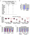
Proper recruitment of pericentrin and γ-tubulin by the hCenexin1-Plk1 complex requires Plk1 function. (A–C), The DT 10 h cells prepared as in Fig. 4A were stained with the indicated antibodies. Recruitment of various centrosomal components to the ectopic hCenexin1 dots were examined among the cells in late G2 or early M phase, the stages where Plk1 is abundantly expressed. Intensities of the recruited signals were categorized from strong (++++) to undetectable (−) (A). The percentage of pericentrin colocalization with the ectopic hCenexin1 dots was quantified in > 300 cells (B). Asterisk denotes weakly recruited pericentrin signals. Bars, standard deviation. (C), EGFP-hCenexin1-expressing HeLa cells or the EGFP-hCenexin1 cells silenced for Plk1 (sh-Plk1) were released from a G1/S block into fresh medium for 10 h. A fraction of the unsilenced cells were treated with BI2536 for 3 h before harvest. To minimize an indirect effect of prolonged Plk1 depletion or inhibition, the total period of release was kept for 10 h and only the prophase cells with condensed chromosomes were analyzed. Bars (red), the averages of colocalization efficiency. Plk1-positive cells (marked in red) indicate a fraction of the sh-Plk1 cells with yet detectable Plk1 signals. (D–E), To examine the effect of Plk1 inhibition or depletion on the localization of hCenexin1, pericentrin, and γ-tubulin to the centrosomes, HeLa cells infected with control sh-Luc or sh-Plk1 lentivirus for 1 day were first arrested by double thymidine treatment. Cells were then released into either nocodazole-containing medium for 12 h to trap the cells in prometaphase (D) or fresh medium for 6 h to enrich the cells in S/G2 (E). BI2536 treatment was carried out 3 h prior to harvest. Bars, standard deviation.
Fig. 6
Fig. 6
S796-dependent hCenexin1 function is important for proper mitotic progression, but not for ninein localization and ciliogenesis. (A–C), HeLa cells expressing the indicated hCenexin1 or hODF2 construct were depleted of endogenous hCenexin1 and hODF2 by si-781 or si-1235. The cells were released from a G1/S block for 10 h and subjected to immunoblotting (A) and immunostaining (B) analyses. Asterisk in (A), a non-specific cross-reacting protein. (C), The cells in (B) exhibiting abnormal chromosome segregation or multipolar spindles were quantified. (D), HeLa cells expressing hCenexin1 or hCenexin1(S796A) were depleted of endogenous hCenexin1 and hODF2. Cells releasing from a G1/S block were subjected to time-lapse microscopy (see Fig. S9). The length of time required for pre- or post-anaphase progression was quantified. Red bars with numbers, the averages of time-length in minute. (E), Asynchronously growing HeLa cells expressing the indicated constructs were depleted of endogenous hCenexin1 and hODF2 as in (A), and then subjected to immunostaining analyses. Since ninein delocalizes from the mitotic centrosomes, ninein configuration was examined among the interphase cells (n > 400). (Right), Representative images of ninein localization with 3+1 or 1+1 configuration are shown. Asterisks indicate the 3+1 configuration with poorly separated ninein dots. (F), hTERT-RPE cells were infected with lentivirus expressing control luciferase (sh-Luc), sh-718 (for depletion of both hCenexin1 and hODF2), or sh-2066 (for depletion of hCenexin1 only), cultured under serum starvation for 2 days, and then immunostained. Samples were quantified to determine the percentages of primary cilia formation (> 400 cells) (Left), the length of primary cilia (> 50 cells) (Middle), and the fluorescence intensities of hCenexin1 signals (> 50 cells) (Right). Error bars, standard deviation. (G–H), hTERT-RPE cells expressing the indicated hCenexin1 or hODF2 constructs were depleted of endogenous hCenexin1 and hODF2 by si-781 or si-1235. Two days after serum starvation, cells were immunostained (G) and quantified in over 500 cells (H). Percentages in (G), efficiency of localization along the axoneme of cilia; Bars in (H), standard deviation; Asterisks in (G) and (H), hCenexin1(1–613) exhibits significantly shorter primary cilia than other constructs.
Fig. 7
Fig. 7
A model illustrating multiple functions of hCenexin1 in both Plk1-independent ninein recruitment and ciliogenesis and Plk1-dependent microtubule nucleation at distinct stages of the cell cycle. (A), In G0/G1 stage of the cell cycle, hCenexin1 plays a critical role in the formation of primary cilia (vertical cylinder). Throughout interphase, hCenexin1 is also required for proper recruitment of ninein to the distal/subdistal appendages of mother centrioles (M), an event that may contribute to proper ciliogenesis. Consistent with a low level of Plk1 expression during interphase, both ninein recruitment and primary cilia formation occur in a Plk1-independent manner. During the late stages of the cell cycle (late G2 and early M), Cdc2 phosphorylates hCenexin1 at S796 and generates a PBD-docking site crucial for Plk1 recruitment to the centrosomes. The formation of the hCenexin1-Plk1 complex is important for proper recruitment of pericentrin, which in turn facilitates γ-tubulin accumulation and centrosome maturation, thus promoting microtubule nucleation. (B), Absence of Cdc2-dependent hCenexin1 phosphorylation at S796 impairs Plk1 recruitment to the centrosomes, which results in reduced levels of recruited pericentrin and γ-tubulin and, therefore, improper microtubule nucleation. Both ninein localization and primary cilia formation at the interphase centrosomes occur normally under these conditions. Cdc2/CycB, B-type cyclin-associated Cdc2 activity; M, mother centrioles, D, daughter centrioles.

References

    1. Barr FA, Sillje HH, Nigg EA. Polo-like kinases and the orchestration of cell division. Nat Rev Mol Cell Biol. 2004;5:429–440. - PubMed
    1. Bellanger S, de Gramont A, Sobczak-Thépot J. Cyclin B2 suppresses mitotic failure and DNA re-replication in human somatic cells knocked down for both cyclins B1 and B2. Oncogene. 2007;26:7175–7184. - PubMed
    1. Brohmann H, Pinnecke S, Hoyer-Fender S. Identification and characterization of new cDNAs encoding outer dense fiber proteins of rat sperm. J Biol Chem. 1997;272:10327–10332. - PubMed
    1. Casenghi M, Meraldi P, Weinhart U, Duncan PI, Korner R, Nigg EA. Polo-like kinase 1 regulates Nlp, a centrosome protein involved in microtubule nucleation. Dev Cell. 2003;5:113–125. - PubMed
    1. de Cárcer G, do Carmo Avides M, Lallena MJ, Glover DM, González C. Requirement of Hsp90 for centrosomal function reflects its regulation of Polo kinase stability. EMBO J. 2001;20:2878–2884. - PMC - PubMed

Publication types

MeSH terms

LinkOut - more resources