. 2009 Apr;16(4):563-75.
doi: 10.1016/j.devcel.2009.02.011.

Linking asymmetric cell division to the terminal differentiation program of postmitotic neurons in C. elegans

Affiliations

Linking asymmetric cell division to the terminal differentiation program of postmitotic neurons in C. elegans

Vincent Bertrand et al. Dev Cell. 2009 Apr.

Abstract

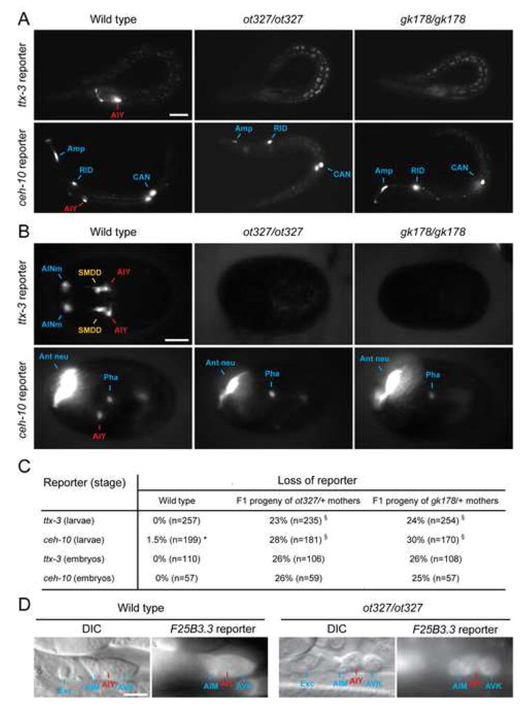
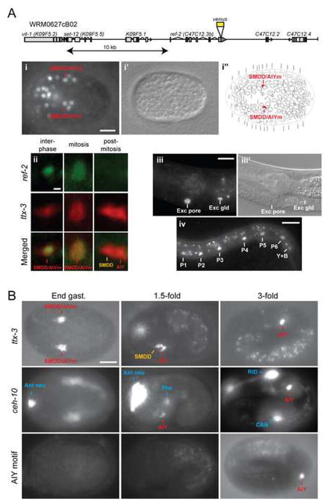
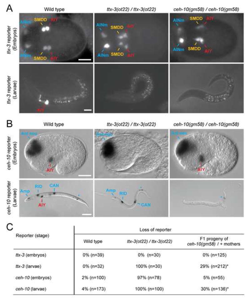
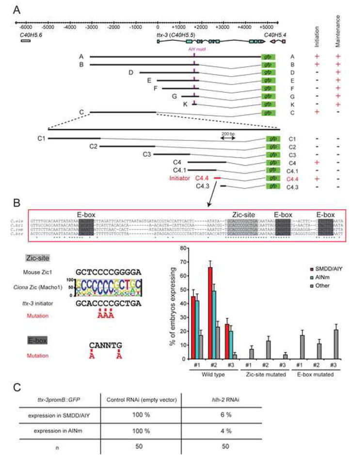
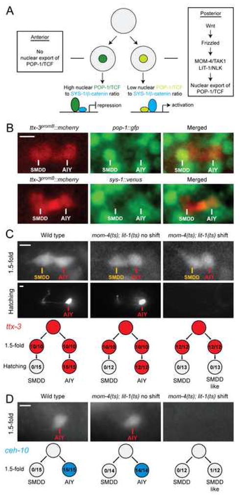
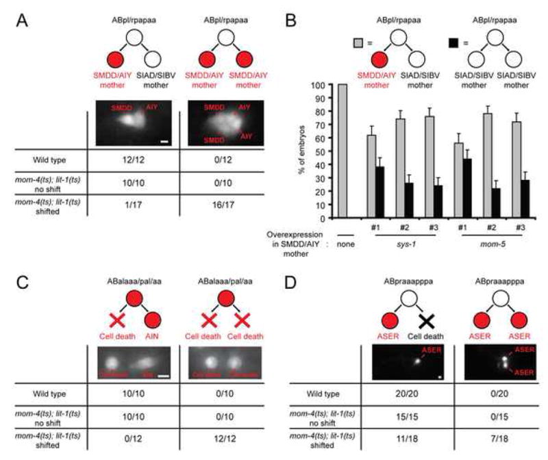
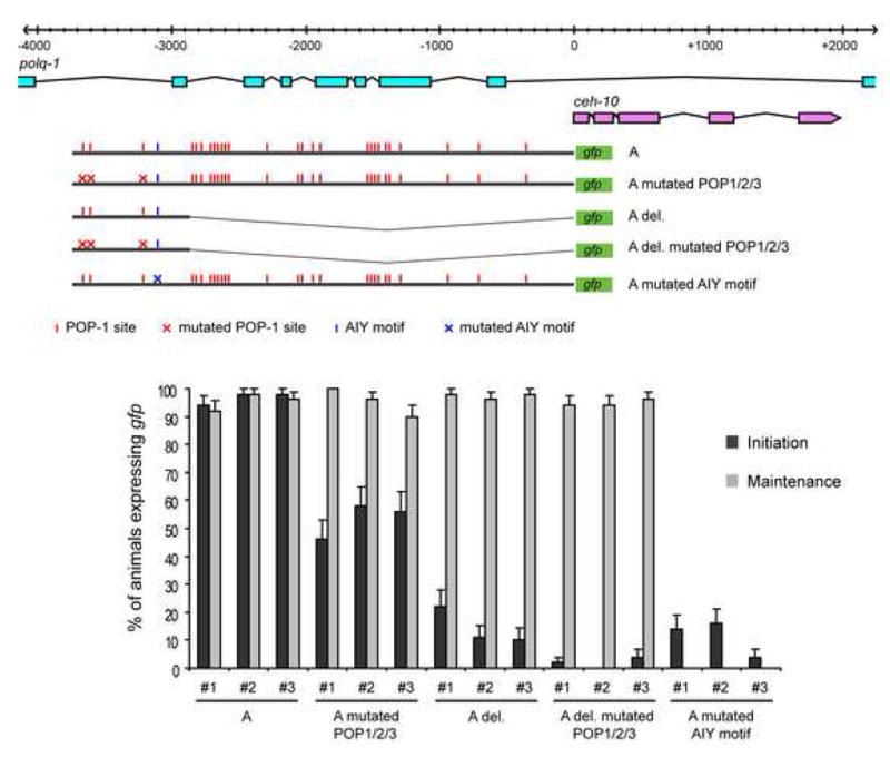
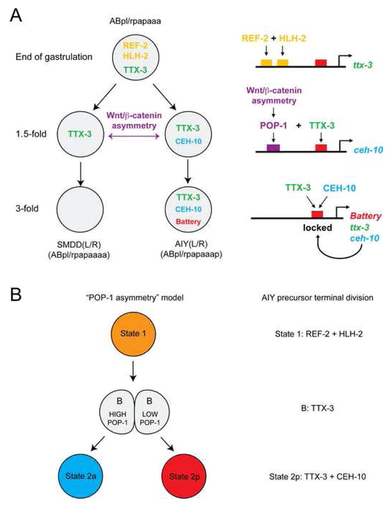
How asymmetric divisions are connected to the terminal differentiation program of neuronal subtypes is poorly understood. In C. elegans, two homeodomain transcription factors, TTX-3 (a LHX2/9 ortholog) and CEH-10 (a CHX10 ortholog), directly activate a large battery of terminal differentiation genes in the cholinergic interneuron AIY. We establish here a transcriptional cascade linking asymmetric division to this differentiation program. A transient lineage-specific input formed by the Zic factor REF-2 and the bHLH factor HLH-2 directly activates ttx-3 expression in the AIY mother. During the terminal division of the AIY mother, an asymmetric Wnt/beta-catenin pathway cooperates with TTX-3 to directly restrict ceh-10 expression to only one of the two daughter cells. TTX-3 and CEH-10 automaintain their expression, thereby locking in the differentiation state. Our study establishes how transient lineage and asymmetric division inputs are integrated and suggests that the Wnt/beta-catenin pathway is widely used to control the identity of neuronal lineages.


Figures

Figure 1
Figure 1. ot327 and gk178 affect the expression of the TTX-3/CEH-10 complex in AIY
(A) In ot327 and gk178 animals, ttx-3 (ttx-3promB::gfp, otIs173) and ceh-10 (ceh-10promA::gfp, lqIs4) expression is lost in AIY at larval stages (lateral view, anterior is left, dorsal is up, Amp: amphid support cell, scale bar = 20 μm). (B) Expression of the same reporters is also lost in the AIY lineage in the embryo (ttx-3: epidermal enclosure, ventral view, anterior is left; ceh-10: 1.5-fold, lateral view, anterior is left, dorsal is up; AINm: AIN mother; Ant neu: anterior neurons; Pha: pharyngeal cell, scale bar = 10 μm). (C) Percentage of animals which have lost reporter expression in the AIY lineage. As ot327 and gk178 are larval lethal, the F1 progeny of heterozygote mothers was analyzed. Around 1/4 of the progeny displays a mutant phenotype, as expected from Mendel’s laws. Embryos were scored between epidermal enclosure and 2-fold for ttx-3, and at 1.5-fold for ceh-10 (*: expression is absent from only one of the two AIY; §: in 3 to 4% of the animals expression is lost in only one of the two AIY). (D) In wild type larvae AIY is located in a group of three neurons just posterior to the excretory cell (Exc) and expressing the pan-neuronal marker F25B3.3 (F25B3.3::dsRed2, otIs173, n=10). In ot327 homozygotes (recognized by the loss of ttx-3promB::gfp expression) a neuron is still present at the position normally occupied by AIY and still expresses F25B3.3::dsRed2 (n=11) (scale bar = 5 μm).
Figure 2
Figure 2. Sequential activation of ref-2, ttx-3, ceh-10 and the AIY motif
(A) Expression pattern of a ref-2::venus rescuing translational fusion gene, observed with the transgenic array otEx3091. (i-i″) Expression in the SMDD/AIY mother (SMDD/AIYm) at the end of gastrulation (ventral view anterior is left, scale bar = 10 μm). (ii) Expression of REF-2::VENUS in the SMDD/AIY lineage (marked by ttx-3promB::mcherry, otIs181) before (interphase), during (mitosis) and after (post-mitosis) cleavage of the SMDD/AIY mother (scale bar = 2 μm). (iii, iii′) Expression in the excretory pore cell (Exc pore) and excretory gland (Exc gld) at L2 larval stage (lateral view, anterior is left, dorsal is up, scale bar = 10 μm). (iv) expression in the P, Y and B-cells at L1 larval stage (lateral view, anterior is left, dorsal is up, scale bar = 20 μm). (B) Comparison of the expression of ttx-3 (ttx-3promB::gfp, mgIs18 or ttx-3promB::mcherry, otEx2644), ceh-10 (ceh-10promA::gfp, lqIs4) and the AIY motif (AIY motif::gfp, otEx1597) at the end of gastrulation (End gast., ventral view, anterior is left), 1.5- and 3-fold (lateral view, anterior is left). At 1.5-fold, the ttx-3 and ceh-10 pictures show colocalization of ttx-3promB::mcherry and ceh-10promA::gfp expression in AIY in the same embryo (scale bar = 10 μm).
Figure 3
Figure 3. Initiation and automaintenance phases of ttx-3 and ceh-10
(A) Effect of ttx-3(ot22) and ceh-10(gm58) alleles on ttx-3 expression (ttx-3promB::gfp, mgIs18) at epidermal enclosure (ventral view, anterior to the left, AINm = AIN mother, scale bar = 10 μm) and larval stage (lateral view, anterior to the left, dorsal to the top, scale bar = 20 μm). (B) Effect of ttx-3(ot22) and ceh-10(gm58) alleles on ceh-10 expression (ceh-10::lacZ, pInt1) at 1.5-fold and larval stage (lateral view, anterior to the left, dorsal to the top, Ant neu: anterior neurons; Amp: amphid support cell; *: variable expression in the posterior intestine, scale bar = 10 μm for 1.5-fold and 50 μm for larva). (C) Percentage of animals which have lost, in the AIY lineage, expression of ttx-3 (ttx-3promB::gfp, mgIs18; embryos were scored between epidermal enclosure and 2-fold) or ceh-10 (ceh-10::lacZ, pInt1; embryos were scored at 1.5-fold). As ceh-10(gm58) is larval lethal, the F1 progeny of heterozygote mothers was analyzed (*: around 1/4 of the progeny displays a mutant phenotype, as expected from Mendel’s laws). A very dim GFP signal was detected in some ttx-3(ot22); ttx-3promB::gfp larvae and in some ceh-10(gm58); ttx-3promB::gfp larvae which may reflect perdurance of GFP signal from earlier expression.
Figure 4
Figure 4. The initiation of ttx-3 expression relies on Zic and bHLH binding sites
(A) Isolation of the cis-regulatory element responsible for the initiation of ttx-3 expression. For each deletion construct the activity in the AIY lineage is presented to the right (initiation: epidermal enclosure to 2-fold, maintenance: 3-fold to adult, purple box: AIY motif, scale in bp, see Figure S2 for quantification). (B) Analysis of the initiator element (C4.4). The upper part presents the alignment of the initiator element sequence between C. elegans (C. ele), C. briggsae (C. bri), C. remanei (C. rem) and C. brenneri (C. bre). The graph presents the percentage of embryos showing expression in the SMDD/AIY lineage (SMDD/AIY, red), AIN lineage (AINm, green) or other lineages (mostly pharynx, grey). The F1 progeny of Rol mothers (positive for the coinjection marker rol-6(d)) was analyzed between epidermal enclosure and 2-fold stage. Three independent lines were scored for each construct (n=100, error bars = standard error to the proportion). (C) Effect of hlh-2 RNAi on the initiation of ttx-3 expression. The table presents the percentage of embryos showing expression of ttx-3promB::gfp (mgIs18) in the SMDD/AIY lineage and AIN lineage during epidermal enclosure after RNAi treatment.
Figure 5
Figure 5. The Wnt/-catenin pathway is involved in the asymmetric division of the SMDD/AIY mother
(A) General diagram of the Wnt/β-catenin asymmetry pathway. Only the components used in this study are presented, for a more complete description of the pathway see Mizumoto and Sawa (2007). Upon stimulation of a Wnt receptor Frizzled, the kinases MOM-4/TAK-1 and LIT-1/NLK are activated in the posterior daughter cell. LIT-1 enters the nucleus and phosphorylates POP-1/TCF promoting its nuclear export. As a consequence POP-1 concentration is low in the posterior nucleus allowing most of the POP-1 proteins to be associated with the limiting coactivator SYS-1/β-catenin and activate transcription, while in the anterior nucleus POP-1 concentration is high, most of the POP-1 proteins are not associated with SYS-1 and therefore repress transcription. (B) Localization of POP-1::GFP (qIs74) or SYS-1::VENUS (qIs95) fusion proteins just after cleavage of the SMDD/AIY mother (epidermal enclosure, ventral view, anterior to the left, SMDD/AIY lineage marked by ttx-3promB::mcherry, otIs181 or otEx2644). (C) Expression of ttx-3 (ttx-3promB::gfp, mgIs18) in wild type or mom-4(ne1539); lit-1(t1512) temperature-sensitive mutants. Embryos were shifted to the restrictive temperature just before cleavage of the SMDD/AIY mother and analyzed at the 1.5-fold stage or at hatching (lateral view, anterior to the left). The results are summed up at the bottom (red = ttx-3 expression, numbers = number of animals showing expression in the cell depicted/total number of animals). (D) Expression of ceh-10 (ceh-10promA::gfp, lqIs4) in wild type or mom-4(ne1539); lit-1(t1512) temperature-sensitive mutants. Embryos were shifted to the restrictive temperature just before cleavage of the SMDD/AIY mother and analyzed at the 1.5-fold stage (lateral view, anterior to the left). The results are summed up at the bottom (blue = ceh-10 expression, numbers = number of embryos showing expression in the cell depicted/total number of embryos). Scale bar for all panels = 2 μm.
Figure 6
Figure 6. The Wnt/-catenin pathway is involved in the asymmetric division of other embryonic neuroblasts
(A) Effect of mom-4(ne1539); lit-1(t1512) temperature-sensitive mutants on the asymmetric division of the ABpl/rpapaa neuroblast. This division generates the SMDD/AIY mother anteriorly and the SIAD/SIBV mother posteriorly. ttx-3 (red) is expressed in the SMDD/AIY mother and the newly born SMDD and AIY but not in the SIAD/SIBV mother or the newly born SIAD and SIBV. Embryos were shifted to the restrictive temperature just before cleavage of the ABpl/rpapaa neuroblast and analyzed at the end of epidermal enclosure (just after the birth of SMDD, AIY, SIAD and SIBV; ventral view, anterior to the left, numbers = number of embryos showing ttx-3promB::gfp, mgIs18 expression in the newly born SMDD and AIY only (left column) or in both the newly born SMDD, AIY and the newly born SIAD, SIBV (right column) over total number of embryos analyzed). (B) Effect of sys-1/-catenin and mom-5/frizzled overexpression on the asymmetric division of the ABpl/rpapaa neuroblast. The graph presents the percentage of embryos showing expression of ttx-3promB::gfp, mgIs18 in the SMDD/AIY lineage but not the SIAD/SIBV lineage (grey) or no expression in those lineages (black). The F1 progeny of control mothers or Rol mothers (positive for the coinjection marker rol-6(d) that marks the sys-1 and mom-5 overexpression arrays) was analyzed between the end of epidermal enclosure and the 1.5-fold stage. Three independent lines were scored for each construct (n>50, error bars = standard error to the proportion). (C) Effect of mom-4(ne1539); lit-1(t1512) temperature-sensitive mutants on the asymmetric division of the AIN mother. This division generates a cell death (rounded shape) and the AIN neuron (elongated shape), both marked by ttx-3promB::gfp expression (red). Although this division is left-right oriented, the daughter that will eventually die is located more anteriorly just after cleavage and displays a higher POP-1 nuclear level than its sister cell AIN. Embryos were shifted to the restrictive temperature just before cleavage of the AIN mother and analyzed at the 1.5-fold stage (lateral view, numbers = number of embryos showing a cell death + neuron pair (left column) or a cell death + cell death pair (right column) over total number of embryos analyzed). (D) Effect of mom-4(ne1539); lit-1(t1512) temperature-sensitive mutants on the asymmetric division of the ASER mother. This division generates the ASER neuron anteriorly (marked by gcy-5::gfp, ntIs1 expression, red) and a cell death posteriorly. Embryos were shifted to the restrictive temperature just before cleavage of the ASER mother and analyzed at hatching (lateral view, anterior to the left, numbers = number of embryos showing only one ASER neuron (left column) or a pair of ASER neurons (right column) over total number of embryos analyzed). Scale bar for all panels = 2 μm.
Figure 7
Figure 7. The initiation of ceh-10 expression relies on TTX-3 and POP-1 binding sites
The upper part depicts the gfp reporter constructs tested relative to the ceh-10 locus (ceh-10 and its cis-regulatory sequences are located in introns of the polymerase gene polq-1 which is oriented in the opposite direction). The graph presents the percentage of animals showing expression in AIY at the 1.5-fold stage (initiation) or early larval stage (maintenance). Transgenic animals were recognized by the presence of reporter gene expression in the CAN lineage, which consistently expresses ceh-10. Three independent lines were scored for each construct (n=50, error bars = standard error to the proportion).
Figure 8
Figure 8. A model for the initiation of the terminal differentiation program of the AIY interneuron
(A) Summary of the regulatory events occurring upon terminal division of the SMDD/AIY mother. Note that arrows between transcription factors and cis-regulatory regions represent likely direct interactions based on the fact that a loss of function of the trans-acting factor is phenocopied by the mutation of its predicted binding site(s) in the respective cis-regulatory region. (B) The terminal division of AIY can be put into a previously proposed theoretical framework (Lin et al., 1998) describing how the “POP-1 asymmetry” can generate specific regulatory states. Note that the putative transcription factor “B” corresponds to TTX-3 in the case of AIY.

Similar articles

Cited by

References

    1. Alper S, Kenyon C. The zinc finger protein REF-2 functions with the Hox genes to inhibit cell fusion in the ventral epidermis of C. elegans. Development. 2002;129:3335–3348. - PubMed
    1. Altun-Gultekin Z, Andachi Y, Tsalik EL, Pilgrim D, Kohara Y, Hobert O. A regulatory cascade of three homeobox genes, ceh-10, ttx-3 and ceh-23, controls cell fate specification of a defined interneuron class in C. elegans. Development. 2001;128:1951–1969. - PubMed
    1. Arata Y, Kouike H, Zhang Y, Herman MA, Okano H, Sawa H. Wnt signaling and a Hox protein cooperatively regulate psa-3/Meis to determine daughter cell fate after asymmetric cell division in C. elegans. Dev Cell. 2006;11:105–115. - PubMed
    1. Aruga J. The role of Zic genes in neural development. Mol Cell Neurosci. 2004;26:205–221. - PubMed
    1. Asbreuk CH, van Schaick HS, Cox JJ, Kromkamp M, Smidt MP, Burbach JP. The homeobox genes Lhx7 and Gbx1 are expressed in the basal forebrain cholinergic system. Neuroscience. 2002;109:287–298. - PubMed

Publication types