. 2009 Jul;83(13):6779-89.
doi: 10.1128/JVI.02443-08. Epub 2009 Apr 22.

Human papillomavirus type 16 (HPV-16) virus-like particle L1-specific CD8+ cytotoxic T lymphocytes (CTLs) are equally effective as E7-specific CD8+ CTLs in killing autologous HPV-16-positive tumor cells in cervical cancer patients: implications for L1 dendritic cell-based therapeutic vaccines

Affiliations

Human papillomavirus type 16 (HPV-16) virus-like particle L1-specific CD8+ cytotoxic T lymphocytes (CTLs) are equally effective as E7-specific CD8+ CTLs in killing autologous HPV-16-positive tumor cells in cervical cancer patients: implications for L1 dendritic cell-based therapeutic vaccines

Stefania Bellone et al. J Virol. 2009 Jul.

Abstract

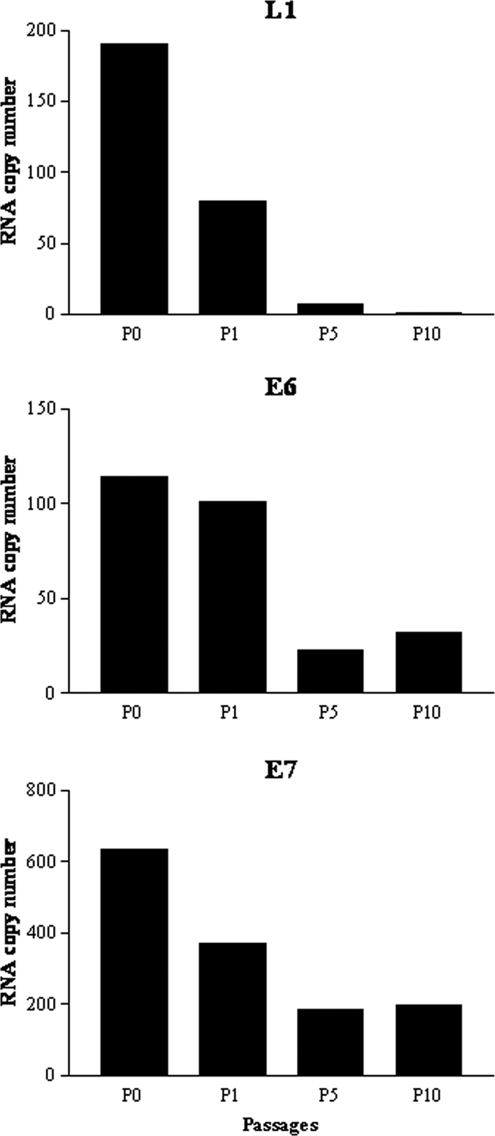
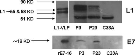
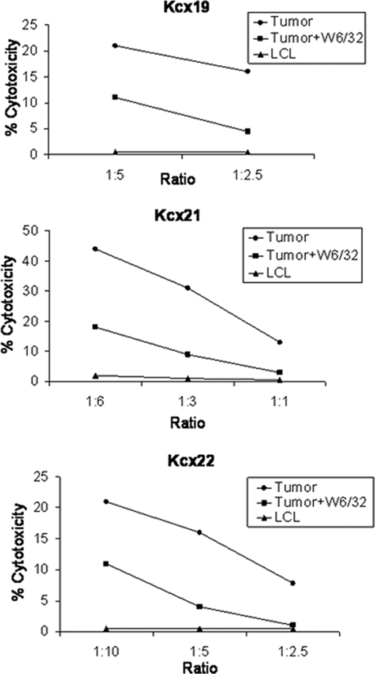
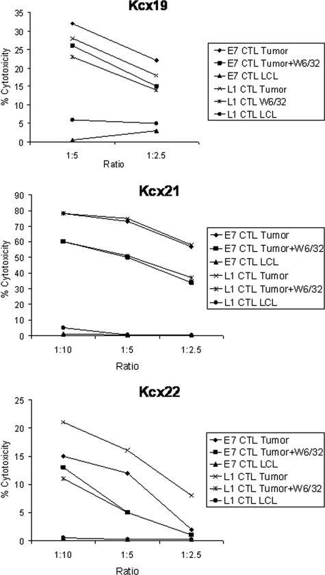
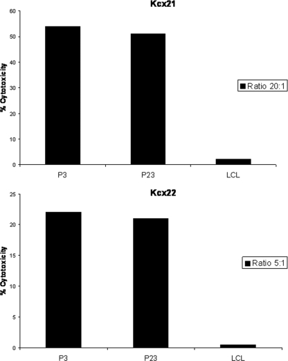
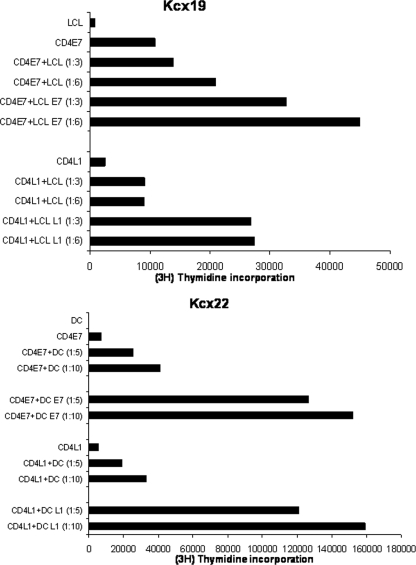
Papillomavirus-like particles (VLPs) based on L1 capsid protein represent a promising prophylactic vaccine against human papillomavirus (HPV) infections. However, cell-mediated immune responses against this antigen are believed to be of limited therapeutic value in established HPV-infected cervical lesions and, for this reason, have not been intensively investigated in cervical cancer patients. In this study we analyzed and quantified by real-time PCR (RT-PCR) the RNA expression levels of E6, E7, and L1 genes in flash-frozen HPV-16 cervical carcinomas. In addition, the kinetics of expression of E6, E7, and L1 in HPV-16-infected primary cell lines established as long-term cultures in vitro was also evaluated at RNA and protein levels. Finally, in order to evaluate the therapeutic potential of L1-specific CD4(+) and CD8(+) T lymphocytes responses in cervical cancer patients, L1 VLP-loaded dendritic cells (DCs) were used to stimulate peripheral blood lymphocytes from cervical cancer patients and such responses were compared to those elicited by the E7 oncoprotein. We show that 22 of 22 (100%) flash-frozen cervical biopsy samples collected from HPV-16-positive cervical cancer patients harbor L1, in addition to E6 and E7 RNA, as detected by RT-PCR. E7 RNA copy number (mean, 176.2) was significantly higher in HPV-16-positive cervical cancers compared to the E6 RNA copy number (mean, 47.3) and the L1 copy number (mean, 58.3) (P < 0.0001 and P < 0.001, respectively). However, no significant differences in expression levels between E6 and L1 were found. Kinetic studies of E6, E7, and L1 RNA and protein expression levels in primary tumors showed a sharp reduction in L1 expression after multiple in vitro passages compared to E6 and E7. Autologous DCs pulsed with HPV-16 VLPs or recombinant full-length E7 elicited strong type 1 L1- and E7-specific responses in CD4(+) and CD8(+) T cells from cervical cancer patients. Importantly, L1 VLP-specific CD8(+) T lymphocytes expressed strong cytolytic activity against autologous tumor cells and were as effective as E7-specific cytotoxic T lymphocytes in lysing naturally HPV-16-infected autologous tumor cells. Taken together, these data demonstrate a consistent expression of L1 in primary cervical tumors and the possibility of inducing effective L1/tumor-specific CD4(+) and CD8(+) T-lymphocyte responses in patients harboring HPV-infected cervical cancer. These results may have important implications for the treatment of patients harboring established HPV-infected lesions with L1 VLPs or combined E7/L1 DC-based vaccinations.


Figures

FIG. 1.
FIG. 1.
Representative qRT-PCR analysis of L1, E6, and E7 expression in a Kcx22 primary HPV-16-positive cell line at different time points of in vitro culture. y axis, fold induction relative to SiHa expression; x axis, each sample from Kcx22 tested for L1, E6, and E7 expression at different time points (i.e., tumors never passed in culture to a new flask = P0, as well as P1, P5, and P10). A clear downward trend of L1 expression to almost undetectable levels over passage number is evident.
FIG. 2.
FIG. 2.
Representative Western blot analysis of L1 (upper panel) and E7 (lower panel) expression in the Kcx22 primary HPV-16-positive cell line at P3 and P23 of in vitro culture. Unlike E7 protein, a visible reduction in L1 protein expression level was consistently noted by comparing early (P3) versus late (P23) in vitro passages in multiple experiments. L1-VLP, recombinant L1 protein; rE7, recombinant E7 protein; C33A, HPV-16-negative control cell line. The double band at approximately ca. 55 to 58 kDa corresponds to posttranslational modification of L1, while the band at ∼51 kDa represents a cellular protein cross-reacting with MAb Camvir-1 (33).
FIG. 3.
FIG. 3.
Tumor-specific HPV-16 L1-specific CD8+ CTL responses induced by L1-pulsed DCs in Kcx19, Kcx21, and Kcx22 patients, measured in a 5-h 51Cr-release assay. Percentage lysis against autologous tumor cells and HLA-identical HPV-16-negative control LCL at different effector/target cell ratio is shown. Anti-HLA class I blocking antibody (W6/32) was used at 50 μg/ml. A representative experiment for each patient is shown.
FIG. 4.
FIG. 4.
Tumor-specific HPV-16 L1-specific CD8+ CTL responses induced by L1-pulsed DCs and tumor-specific HPV-16 E7-specific CD8+ CTL responses induced by E7-pulsed DCs in Kcx19, Kcx21, and Kcx22 patients, as measured in a 5-h 51Cr-release assay. The percent lysis against autologous tumor cells and HLA-identical HPV-16-negative control LCL at different effector/target cell ratio is shown. Anti-HLA class I blocking antibody (W6/32) was used at 50 μg/ml. A representative experiment for each patient is shown.
FIG. 5.
FIG. 5.
Tumor-specific HPV-16 L1-specific CD8+ CTL responses induced by L1-pulsed DCs in Kcx21 and Kcx22 patients, measured in a 5-h 51Cr-release assay. The percent lysis against autologous tumor cells at early passages (i.e., P3) and late passages (i.e., P23) and HLA-identical HPV-16-negative control LCL at different effector/target cell ratios is shown.
FIG. 6.
FIG. 6.
(Upper panel) CD4+ T-cell proliferation in response to stimulation with HPV E7- or L1-pulsed autologous irradiated LCL (LCL/E7 and LCL/L1) at different effector/target cell ratios in patient Kcx19. (Lower panel) CD4+ T-cell proliferation in response to stimulation with HPV E7- or L1-pulsed autologous DCs (DC/E7 and DC/L1) at different effector/target cell ratio in patient Kcx22. [3H]thymidine (1 mCi/well) was added to each well for at least 16 h of the assay, and proliferation was determined by measuring the [3H]thymidine incorporation. The difference in mean proliferations determined by measuring the [3H]thymidine incorporation in the presence of LCL/E7-DC/E7 or LCL/L1-DC/L1 compared to that in the presence of LCL or DC controls is significant at P < 0.01 as determined by the Student t test for both patients. In crossing experiments, no proliferation was detected in L1 VLP or E7-specific CD4+ T cells when autologous LCL or DCs were loaded with full-length E7 (instead of L1) or L1 (instead of E7) were used as stimulators.

Similar articles

Cited by

References

    1. Aiba, S., M. Rokugo, and H. Tagami. 1986. Immunohistologic analysis of the phenomenon of spontaneous regression of numerous flat warts. Cancer 581246-1251. - PubMed
    1. Banchereau, J., and R. M. Steinman. 1998. Dendritic cells and the control of immunity. Nature 392245-252. - PubMed
    1. Banerjee, D. K., M. V. Dhodapkar, E. Matayeva, R. M. Steinman, and K. M. Dhodapkar. 2006. Expansion of FOXP3high regulatory T cells by human dendritic cells (DCs) in vitro and after injection of cytokine-matured DCs in myeloma patients. Blood 1082655-2661. - PMC - PubMed
    1. Bosch, F. X., A. Lorincz, N. Munoz, C. J. Meijer, and K. V. Shah. 2002. The causal relation between human papillomavirus and cervical cancer. J. Clin. Pathol. 55244-265. - PMC - PubMed
    1. Coleman, N., H. D. Birley, A. M. Renton, N. F. Hanna, B. K. Ryait, M. Byrne, D. Taylor-Robinson, and M. A. Stanley. 1994. Immunological events in regressing genital warts. Am. J. Clin. Pathol. 102768-774. - PubMed

Publication types

MeSH terms

Substances