. 2009 Jul;83(13):6957-62.
doi: 10.1128/JVI.00254-09. Epub 2009 Apr 22.

Detection of nonstructural protein 6 in murine coronavirus-infected cells and analysis of the transmembrane topology by using bioinformatics and molecular approaches

Affiliations

Detection of nonstructural protein 6 in murine coronavirus-infected cells and analysis of the transmembrane topology by using bioinformatics and molecular approaches

Surendranath Baliji et al. J Virol. 2009 Jul.

Abstract

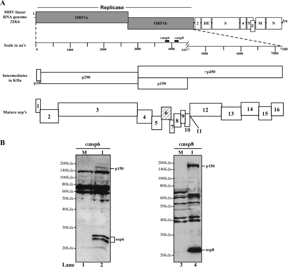
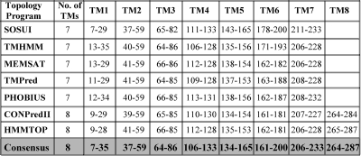
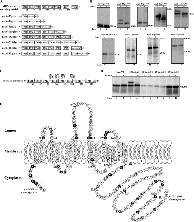
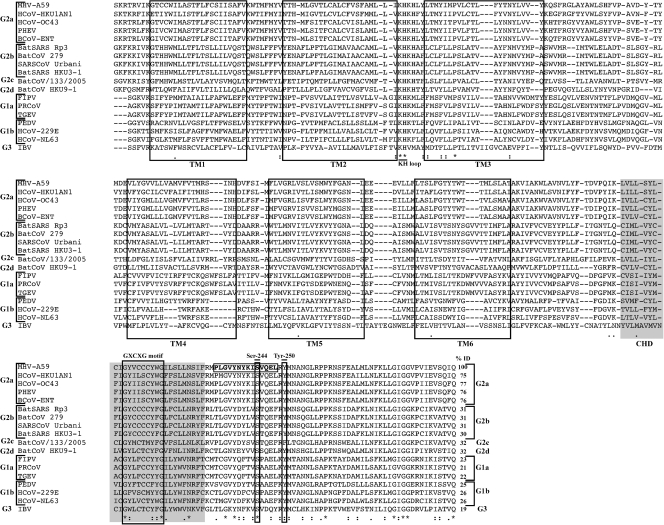
Coronaviruses encode large replicase polyproteins which are proteolytically processed by viral proteases to generate mature nonstructural proteins (nsps) that form the viral replication complex. Mouse hepatitis virus (MHV) replicase products nsp3, nsp4, and nsp6 are predicted to act as membrane anchors during assembly of the viral replication complexes. We report the first antibody-mediated Western blot detection of nsp6 from MHV-infected cells. The nsp6-specific peptide antiserum detected the replicase intermediate p150 (nsp4 to nsp11) and two nsp6 products of approximately 23 and 25 kDa. Analysis of nsp6 transmembrane topology revealed six membrane-spanning segments and a conserved hydrophobic domain in the C-terminal cytosolic tail.


Figures

FIG. 1.
FIG. 1.
Schematic diagram of MHV RNA genome, indicating the proteolytic processing scheme of the replicase polyprotein and Western blot detection of MHV nsp6. (A) MHV-A59 linear RNA genome with the canonical representation of replicase, structural, and accessory genes. The replicase polyprotein intermediates and mature nsps generated during processing are depicted. The mature nsp6 replicase protein (hatched box) and the antibodies used to detect nsp6 and nsp8 (solid black boxes) are indicated. aa's, amino acids. (B) Western blot analysis of nsp6. Whole-cell lysates were prepared from mock-infected (M) and MHV-infected (I) HeLa-MHVR cells, and the lysates were separated by 12.5% SDS-PAGE. Products were detected by probing with nsp6- or nsp8-specific antibodies.
FIG. 2.
FIG. 2.
Summary of TM predictions for MHV nsp6 obtained from membrane topology bioinformatics tools. The nsp6 amino acid sequence (amino acids 3637 to 3923 in the MHV A59 genome are numbered 1 to 287 for simplicity) was analyzed for TM-spanning domains by the use of various bioinformatics tools, and the residue numbers with predicted TM domains are displayed. The consensus TM topology of MHV nsp6 used as a basis for the topology experiments is depicted at the bottom row (shaded in gray).
FIG. 3.
FIG. 3.
Determining the topology of nsp6 by the use of EGFPglyc and insertion of glycosylation consensus sites. (A) Schematics of a working topology model of MHV nsp6 (obtained from our consensus experiments) and nsp6-EGFPglycV5 fusion constructs generated for endo H assay. (B) Metabolic labeling and endo H treatment of nsp6-EGFPglycV5 fusion proteins. The nsp6-EGFPglycV5 fusion proteins expressed in transfected BsrT7 cells were radiolabeled from 20 to 22 h posttransfection, and then cell lysates were subjected to immunoprecipitation with V5 antibody, treated with endo H or left untreated, separated by 12.5% SDS-PAGE, and analyzed by autoradiography. (C) Map of plasmid DNA construct showing the sites of inserted glycosylation acceptor consensus sequences (NXS). The locations of glycosylation insertion in the nsp6-V5 construct are represented, with the amino acid number at the site of insertion. (D) Metabolic labeling and endo H analysis of glycosylation sequence insertion expression constructs of nsp6-V5. The plasmid DNAs (iNsp6-V5 constructs) were transfected and analyzed as described for panel B. (E) MHV-A59 nsp6 topology model, summarizing the results of EGFPglycV5 and glycosylation sequence insertion experiments. Amino acid positions indicated by the symbol “Y” were glycosylated and were positive by endo H assay, whereas those positions tested but found not glycosylated and negative by endo H assay are depicted by solid black horizontal lines. The inserted glycosylation acceptor sequence positions precede the letter i. Selected charged residues are shown in white characters on a black background. K, lysine residues; R, arginine residues; E, glutamic acid residues.
FIG. 4.
FIG. 4.
Multiple sequence alignment (MSA) and percent sequence identity of coronavirus nsp6. The nsp6 amino acid sequences of 18 different coronaviruses were obtained from PATRIC (http://patric.vbi.vt.edu/) and aligned using MUSCLE and ClustalW software. The experimentally determined TM domains of MHV-A59 nsp6 were used as a reference for alignment. Unshaded boxes indicate the conserved TM domains that aligned with other coronavirus nsp6 sequences; the conserved hydrophobic domain (CHD) predicted by all the topology programs is indicated by gray shading. The residues of the peptide against which the nsp6 antibody was raised are boxed, with residue designations shown in boldface. Putative sites for palmitoylation (cysteine residue[s]) within the GXCXG motif) and phosphorylation (serine 244 and tyrosine 250 in MHV-A59 nsp6) are indicated. Percent identity (% ID) values are indicated. In MSA, the following notations were used: asterisk indicate invariant amino acids, colons indicate highly similar amino acids, and dots indicate similar amino acids. HCoV, human coronavirus; PHEV, porcine hemagglutinating encephalomyelitis virus; BCoV, bovine coronavirus; BatSARS, bat severe acute respiratory syndrome coronavirus; BatCoV, bat coronavirus; SARSCoV, severe acute respiratory syndrome coronavirus; FIPV, feline infectious peritonitis virus; PRCoV, porcine respiratory coronavirus; TGEV, transmissible gastroenteritis virus; PEDV, porcine epidemic diarrhea virus; IBV, infectious bronchitis virus.

Similar articles

Cited by

References

    1. Angeles Juanes, M., J. C. Igual, and M. C. Bano. 2008. Membrane topology and post-translational modification of the Saccharomyces cerevisiae essential protein Rot1. Yeast 2593-106. - PubMed
    1. Baker, S. C., and M. R. Denison. 2008. Cell biology of nidovirus replication complexes, p. 103-113. In S. Perlman, T. Gallagher, and E. J. Snijder (ed.), Nidoviruses. ASM Press, Washington, DC.
    1. Baker, S. C., C. K. Shieh, L. H. Soe, M. F. Chang, D. M. Vannier, and M. M. Lai. 1989. Identification of a domain required for autoproteolytic cleavage of murine coronavirus gene A polyprotein. J. Virol. 633693-3699. - PMC - PubMed
    1. Bonilla, P. J., S. A. Hughes, and S. R. Weiss. 1997. Characterization of a second cleavage site and demonstration of activity in trans by the papain-like proteinase of the murine coronavirus mouse hepatitis virus strain A59. J. Virol. 71900-909. - PMC - PubMed
    1. Bost, A. G., R. H. Carnahan, X. T. Lu, and M. R. Denison. 2000. Four proteins processed from the replicase gene polyprotein of mouse hepatitis virus colocalize in the cell periphery and adjacent to sites of virion assembly. J. Virol. 743379-3387. - PMC - PubMed

Publication types

Substances

LinkOut - more resources