

Brain polarization enhances the formation and retention of motor memories
- PMID: 19386757
- PMCID: PMC2712265
- DOI: 10.1152/jn.00184.2009
Brain polarization enhances the formation and retention of motor memories
Abstract
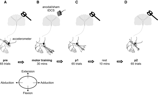
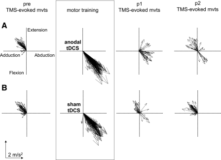
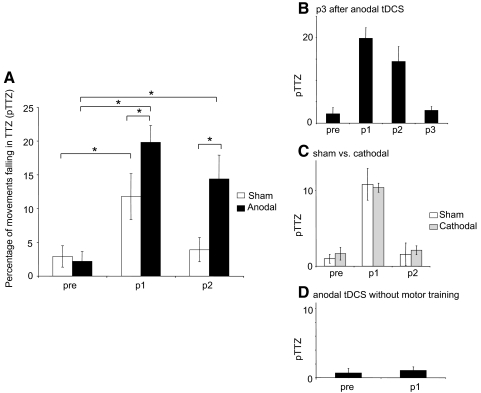
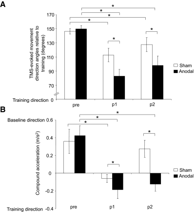
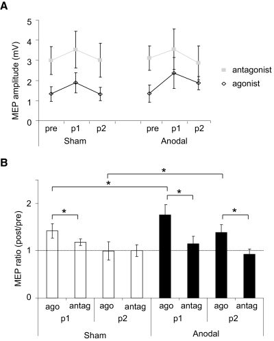
One of the first steps in the acquisition of a new motor skill is the formation of motor memories. Here we tested the capacity of transcranial DC stimulation (tDCS) applied over the motor cortex during motor practice to increase motor memory formation and retention. Nine healthy individuals underwent a crossover transcranial magnetic stimulation (TMS) study designed to test motor memory formation resulting from training. Anodal tDCS elicited an increase in the magnitude and duration of motor memories in a polarity-specific manner, as reflected by changes in the kinematic characteristics of TMS-evoked movements after anodal, but not cathodal or sham stimulation. This effect was present only when training and stimulation were associated and mediated by a differential modulation of corticomotor excitability of the involved muscles. These results indicate that anodal brain polarization can enhance the initial formation and retention of a new motor memory resulting from training. These processes may be the underlying mechanisms by which tDCS enhances motor learning.
Figures
Similar articles
-
Induction of cortical plasticity and improved motor performance following unilateral and bilateral transcranial direct current stimulation of the primary motor cortex.BMC Neurosci. 2013 Jul 1;14:64. doi: 10.1186/1471-2202-14-64. BMC Neurosci. 2013. PMID: 23815634 Free PMC article. Clinical Trial.
-
The effects of bilateral M1 anodal tDCS on corticomotor excitability and acquisition the of a bimanual videogame skill.Neuroscience. 2025 Mar 5;568:231-239. doi: 10.1016/j.neuroscience.2025.01.028. Epub 2025 Jan 19. Neuroscience. 2025. PMID: 39837365
-
The effects of combined repetitive transcranial magnetic stimulation and transcranial direct current stimulation on motor function in patients with stroke.Restor Neurol Neurosci. 2016 Nov 22;34(6):915-923. doi: 10.3233/RNN-160654. Restor Neurol Neurosci. 2016. PMID: 27689549 Clinical Trial.
-
Corticomotor excitability induced by anodal transcranial direct current stimulation with and without non-exhaustive movement.Brain Res. 2013 Sep 5;1529:83-91. doi: 10.1016/j.brainres.2013.07.026. Epub 2013 Jul 24. Brain Res. 2013. PMID: 23891715
-
Modulation of motor performance and motor learning by transcranial direct current stimulation.Curr Opin Neurol. 2011 Dec;24(6):590-6. doi: 10.1097/WCO.0b013e32834c3db0. Curr Opin Neurol. 2011. PMID: 21968548 Review.
Cited by
-
Anodal tDCS over the Primary Motor Cortex Facilitates Long-Term Memory Formation Reflecting Use-Dependent Plasticity.PLoS One. 2015 May 21;10(5):e0127270. doi: 10.1371/journal.pone.0127270. eCollection 2015. PLoS One. 2015. PMID: 25996937 Free PMC article.
-
Probing for hemispheric specialization for motor skill learning: a transcranial direct current stimulation study.J Neurophysiol. 2011 Aug;106(2):652-61. doi: 10.1152/jn.00210.2011. Epub 2011 May 25. J Neurophysiol. 2011. PMID: 21613597 Free PMC article.
-
Contributions of the motor cortex to adaptive control of reaching depend on the perturbation schedule.Cereb Cortex. 2011 Jul;21(7):1475-84. doi: 10.1093/cercor/bhq192. Epub 2010 Dec 3. Cereb Cortex. 2011. PMID: 21131448 Free PMC article.
-
Disrupting the ventral premotor cortex interferes with the contribution of action observation to use-dependent plasticity.J Cogn Neurosci. 2011 Dec;23(12):3757-66. doi: 10.1162/jocn_a_00051. Epub 2011 May 12. J Cogn Neurosci. 2011. PMID: 21563888 Free PMC article. Clinical Trial.
-
Transspinal stimulation increases motoneuron output of multiple segments in human spinal cord injury.PLoS One. 2019 Mar 7;14(3):e0213696. doi: 10.1371/journal.pone.0213696. eCollection 2019. PLoS One. 2019. PMID: 30845251 Free PMC article.
References
-
- Antal A, Nitsche MA, Kincses TZ, Kruse W, Hoffmann KP, Paulus W. Facilitation of visuo-motor learning by transcranial direct current stimulation of the motor and extrastriate visual areas in humans. Eur J Neurosci 19: 2888–2892, 2004. - PubMed
-
- Boggio PS, Alonso-Alonso M, Mansur CG, Rigonatti SP, Schlaug G, Pascual-Leone A, Fregni F. Hand function improvement with low-frequency repetitive transcranial magnetic stimulation of the unaffected hemisphere in a severe case of stroke. Am J Phys Med Rehabil 85: 927–930, 2006. - PubMed
-
- Butefisch CM, Davis BC, Sawaki L, Waldvogel D, Classen J, Kopylev L, Cohen LG. Modulation of use-dependent plasticity by d-amphetamine. Ann Neurol 51: 59–68, 2002. - PubMed
-
- Butefisch CM, Khurana V, Kopylev L, Cohen LG. Enhancing encoding of a motor memory in the primary motor cortex by cortical stimulation. J Neurophysiol 91: 2110–2116, 2004. - PubMed
Publication types
MeSH terms
Grants and funding
LinkOut - more resources
Full Text Sources
Medical