. 2009 Jun;20(12):2864-73.
doi: 10.1091/mbc.e09-02-0168. Epub 2009 Apr 22.

Humanin inhibits neuronal cell death by interacting with a cytokine receptor complex or complexes involving CNTF receptor alpha/WSX-1/gp130

Affiliations

Humanin inhibits neuronal cell death by interacting with a cytokine receptor complex or complexes involving CNTF receptor alpha/WSX-1/gp130

Yuichi Hashimoto et al. Mol Biol Cell. 2009 Jun.

Abstract

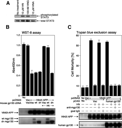
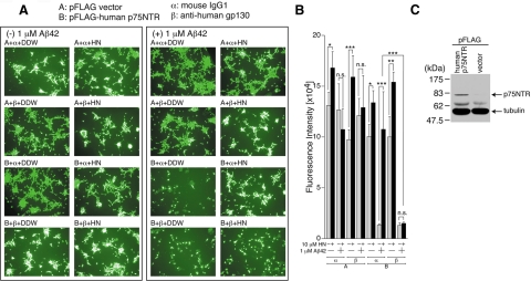
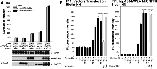
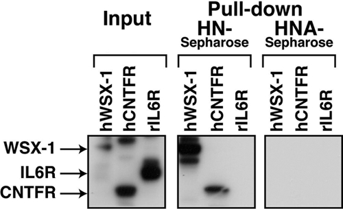
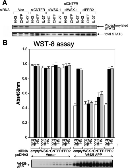
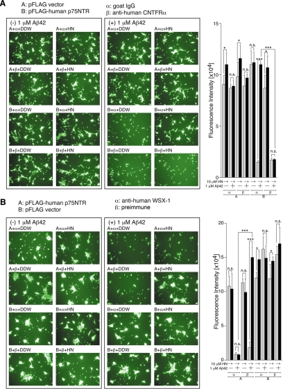
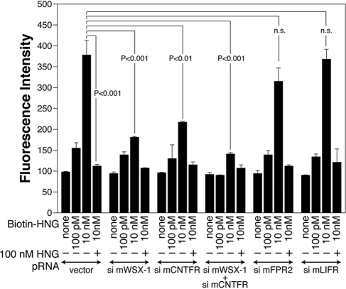
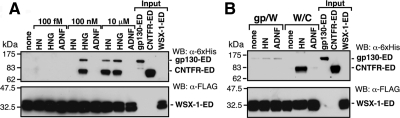
Humanin (HN) inhibits neuronal death induced by various Alzheimer's disease (AD)-related insults via an unknown receptor on cell membranes. Our earlier study indicated that the activation of STAT3 was essential for HN-induced neuroprotection, suggesting that the HN receptor may belong to the cytokine receptor family. In this study, a series of loss-of-function tests indicated that gp130, the common subunit of receptors belonging to the IL-6 receptor family, was essential for HN-induced neuroprotection. Overexpression of ciliary neurotrophic factor receptor alpha (CNTFR) and/or the IL-27 receptor subunit, WSX-1, but not that of any other tested gp130-related receptor subunit, up-regulated HN binding to neuronal cells, whereas siRNA-mediated knockdown of endogenous CNTFR and/or WSX-1 reduced it. These results suggest that both CNTFR and WSX-1 may be also involved in HN binding to cells. Consistent with these results, loss-of-functions of CNTFR or WSX-1 in neuronal cells nullified their responsiveness to HN-mediated protection. In vitro-reconstituted binding assays showed that HN, but not the other control peptide, induced the hetero-oligomerization of CNTFR, WSX-1, and gp130. Together, these results indicate that HN protects neurons by binding to a complex or complexes involving CNTFR/WSX-1/gp130.


Figures

Figure 1.
Figure 1.
gp130 is essential for HN-mediated protection against V642I-APP-induced death of F11 neurohybrid cells. (A) F11 cells, incubated in Ham's F12 medium containing N2 supplement without FBS for 48 h, were treated with 10 μM HN, 100 nM HNG, or 10 μM HNA at 37°C for 15 min and then harvested for immunoblot analysis with the phosphoSTAT3 (Tyr705) and total STAT3 antibodies. (B) F11 cells were transfected with 1.0 μg of the pcDNA3 vector (Vec) or pcDNA3-V642I-APP in association with 1.0 μg of the pCAG vector, pCAG-wild-type mouse gp130 (abbreviated wt), or pCAG-mouse gp130 extracellular domain plus transmembrane domain (dn). Death was induced by the method I in the presence or absence of HN (10 μM). Cell viability was determined with WST-8 assay at 72 h after transfection. Immunoblot analysis was done with an antibody to APP and gp130. Overexpression of wt-gp130 resulted in appearance of a degraded (or misfolded) gp130 product in addition to wt-gp130. (C) F11 cells were transfected with 1.0 μg of the pcDNA3 vector (Vec) or pcDNA3-V642I-APP in association with 1.0 μg of the pCAG vector (Vec) or pCAG-human wt-gp130, as indicated in B. HN (10 μM) and 10 μg of a neutralizing anti-mouse gp130 antibody (R&D Systems) or normal goat IgG were added at 5 h after the onset of transfection. Cell mortality was determined by trypan blue exclusion assay at 72 h after the onset of transfection. Immunoblot analysis was done with antibodies to APP and gp130 (Santa Cruz Biotechnology, C-20).
Figure 2.
Figure 2.
gp130 is essential for HN-mediated protection against Aβ42-induced death of SH-SY5Y cells expressing p75NTR. Human SH-SY5Y cells, transfected with 1.0 μg of the pFLAG vector (Vec) or pFLAG-human p75NTR, were replated onto 96-well dished at 3 h after the onset of transfection. At 24 h after the start of transfection, they were coincubated with 1 μM Aβ42 together with or without 10 μM HN in the presence of 1 μL of 1 mg/ml neutralizing mouse mAb to human gp130 or control mouse IgG. At 48 h after the start of transfection, representative microscopic pictures of the calcein-stained cells were taken (A) and cell viability was determined with calcein-staining assays (B). Immunoblot analysis was done with an antibody to FLAG for the detection of p75NTR-FLAG (C). DDW: distilled water. * p < 0.05; ** p < 0.01; *** p < 0.001; n.s., not significant.
Figure 3.
Figure 3.
Enforced expression of CNTFR or WSX-1 in F11 cells increases the association between HN and F11 cells. (A) F11 cells were cotransfected with pEF1/MycHis-mouse CREME9 (0.5 μg) plus the pcDNA3 vector (0.5 μg) plus pCAG-human gp130 (1.0 μg), pEF1/MycHis-human WSX-1 (0.5 μg) plus the pcDNA3 vector (0.5 μg) plus pCAG-human gp130 (1.0 μg), the pEF1/MycHis vector (0.5 μg) plus pcDNA3.1/GS-human CNTFR (0.5 μg) plus pCAG-human gp130 (1.0 μg), or pEF1/MycHis-human WSX-1 (0.5 μg) plus pcDNA3.1/GS-human CNTFR (0.5 μg) plus pCAG-human gp130 (1.0 μg), for 3 h in the absence of serum. The cells were incubated in HF-18% thereafter. At 24 h after the onset of transfection, cells were replated onto poly-l-lysine–coated 96-well plates (7 × 10³ cells/well). At 36 h after the start of transfection, they were used for immunofluorescence-based binding assay with 10 μM biotin-HN or 10 nM biotin-HNG. Immunoblot analysis was done with a mixture of an anti-human gp130 antibody and an anti-myc mAb (CREME9 and WSX-1), or an anti-V5 mAb (CNTFR). (B) F11 cells, cotransfected with 0.25 μg of the pcDNA3.1/GS vector, 0.25 μg of the pEF1/MycHis vector, and 0.5 μg of the pCAG vector (left panel) or F11 cells, cotransfected with 0.25 μg of pcDNA3.1/GS-human CNTFR, 0.25 μg of pEF1/MycHis-human WSX-1, and 0.5 μg of pCAG-human gp130 (right panel), were replated onto poly-l-lysine–coated 96-well plates at 24 h after transfection. After 12 h of incubation, they were used for immunofluorescence-based binding assay with stepwise increasing concentrations of biotin-HN in the presence or absence of 100 μM of unlabeled HN or HNA as a competitor.
Figure 4.
Figure 4.
HN binds to WSX-1 and CNTFR. F11 cells were transfected with 0.5 μg of pEF1/MycHis-rat IL-6R, pcDNA3.1/GS-human CNTFR, or pEF1/MycHis-human WSX-1. At 48 h after transfection, cells were harvested for pulldown assays with HN or HNA-conjugated Sepharose4B. Precipitants were subjected to immunoblot analysis with a mixture of an anti-myc mAb to detect myc-tagged rat IL-6Rα and human WSX-1, and an anti-V5 mAb to detect V5-tagged human CNTFR.
Figure 5.
Figure 5.
WSX-1 and CNTFR are essential for HN activity in F11 cells. (A) F11 cells were transfected with 0.5 μg of the pRNA-U6.1/Shuttle vector (indicated Vec) or the vector encoding mCNTFR siRNA, mWSX-1 siRNA, or mFPR2 siRNA. Cells were thereafter incubated in HF-18%. In some experiments, F11 cells were cotransfected with 0.5 μg of mCNTFR siRNA- and 0.5 μg of mWSX-1 siRNA-encoding vectors. Total amounts of transfected vectors were kept at 1.0 μg by the addition of appropriate amounts of the backbone vector. At 24 h after the onset of transfection, culture media were replaced by Ham's F12 media containing N2 supplement without FBS. At 72 h after the onset of transfection, cells were incubated with 100 nM HNG, 100 ng/ml human CNTF, or 1 μM human IL-27 at 37°C for 15 min and then harvested for immunoblot analysis with the anti-phosphoSTAT3 (Tyr705) and anti-total STAT3 antibodies. (B) F11 cells were cotransfected with 0.5 μg of the pcDNA3 vector (Vector) or pcDNA3-V642I APP together with 0.5 μg of the pRNA-U6.1/Shuttle vector (indicated empty) or a vector encoding mWSX-1 siRNA, mouse CNTFR siRNA, or mFPR2 siRNA. Cell death was induced by the method II with or without 10 μM HN or 10 nM HNG. WST-8 assays were performed at 72 h after the onset of transfection. Immunoblot analysis was done with the antibody to APP (bottom panel).
Figure 6.
Figure 6.
WSX-1 and CNTFR are essential for HN-mediated protection against Aβ42-induced death of SH-SY5Y cells expressing p75NTR. SH-SY5Y cells, transfected with 1.0 μg of the pFLAG vector (Vec) or pFLAG-human p75NTR, were replated onto 96-well dishes at 3 h after transfection. At 24 h after the start of transfection, they were coincubated with 1 μM Aβ42 together with or without 10 μM HN. One microliter of 1 mg/ml neutralizing goat polyclonal antibody mAb to human CNTFR or control goat IgG was added in A, and 1 μL of rabbit polyclonal antibody to the N-terminal 16 peptide of human WSX-1 or preimmune serum was added in B. At 48 h after the start of transfection, the cell viability was determined with calcein-staining assays. Microscopic pictures of the cells were also taken. DDW, distilled water. * p < 0.05; ** p < 0.01; *** p < 0.001; n.s., not significant.
Figure 7.
Figure 7.
Knockdown of WSX-1 and/or CNTFR reduces the association between HNG and F11 cells. F11 cells were transfected with 0.5 μg of the pRNA-U6.1/Shuttle vector or a vector encoding mWSX-1 siRNA, mCNTFR siRNA, mFPR2 siRNA, or mLIFR siRNA 3 h in the absence of serum and were thereafter incubated with HF-18%. In an experiment, 0.5 μg of mWSX-1 siRNA and mCNTFR siRNA-encoding vectors were cotransfected. Total amounts of transfected vectors were kept at 1.0 μg by the addition of appropriate amounts of the vector. At 24 h after transfection, cells were replated to poly-l-lysine–coated 96-well plates. At 36 h after the start of transfection, they were used for immunofluorescence-based HN-binding assay with indicated concentrations of biotin-HNG together with or without 100 nM of unlabeled HNG.
Figure 8.
Figure 8.
Complex formation of the HN receptor subunits. (A and B) Human CNTFR-ED-Fc-6xHis (CNTFR- ED; 1 μg) and/or mouse gp130-ED-Fc-6xHis (gp130-ED; 1 μg) were coincubated with mouse WSX-1-ED-FLAG (WSX-1-ED; estimated to be 5 μg) beforehand immobilized onto M2 anti-FLAG antibody-conjugated agarose in 100 μl PBS containing 1% Brij 96 for 6 h at 37°C in the presence or absence of indicated concentrations of HN, HNG, or ADNF (A) or in the presence or absence of 10 μM HN or ADNF (B). Washed precipitates were then subjected to immunoblot analysis with anti-6xHis antibody (for CNTFR-ED and gp130-ED) or anti-FLAG (M2) antibody (for WSX-1-ED-FLAG). CNTFR-ED (0.5 μg), gp130-ED (0.5 μg), and WSX-1-ED-FLAG (2.5 μg) immobilized onto M2 anti-FLAG antibody-conjugated agarose were separately subjected to immunoblot analysis as inputs.

Similar articles

Cited by

References

    1. Benaki D., Zikos C., Evangelou A., Livaniou E., Vlassi M., Mikros E., Pelecanou M. Solution structure of humanin, a peptide against Alzheimer's disease-related neurotoxicity. Biochem. Biophys. Res. Commun. 2005;329:152–160. - PubMed
    1. Bozyczko-Coyne D., McKenna B. W., Connors T. J., Neff N. T. A rapid fluorometric assay to measure neuronal survival in vitro. J. Neurosci. Methods. 1993;50:205–216. - PubMed
    1. Boulanger M. J., Garcia K. C. Shared cytokine signaling receptors: structural insights from the gp130 system. Adv. Protein Chem. 2004;68:107–146. - PubMed
    1. Boulay J-L, O'Shea J. J., Paul W. E. Molecular phylogeny within type I cytokines and their cognate receptors. Immunity. 2003;19:159–163. - PubMed
    1. Caricasole A., Bruno V., Cappuccio I., Melchiorri D., Copani A., Nicoletti F. A novel rat gene encoding a Humanin-like peptide endowed with broad neuroprotective activity. FASEB J. 2002;16:1331–1333. - PubMed

Publication types

MeSH terms

Substances

LinkOut - more resources