Satellite cell-mediated angiogenesis in vitro coincides with a functional hypoxia-inducible factor pathway
- PMID: 19386789
- PMCID: PMC2692418
- DOI: 10.1152/ajpcell.00391.2008
Satellite cell-mediated angiogenesis in vitro coincides with a functional hypoxia-inducible factor pathway
Abstract
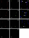
Muscle regeneration involves the coordination of myogenesis and revascularization to restore proper muscle function. Myogenesis is driven by resident stem cells termed satellite cells (SC), whereas angiogenesis arises from endothelial cells and perivascular cells of preexisting vascular segments and the collateral vasculature. Communication between myogenic and angiogenic cells seems plausible, especially given the number of growth factors produced by SC. To characterize these interactions, we developed an in vitro coculture model composed of rat skeletal muscle SC and microvascular fragments (MVF). In this system, isolated epididymal MVF suspended in collagen gel are cultured over a rat SC monolayer culture. In the presence of SC, MVF exhibit greater indices of angiogenesis than MVF cultured alone. A positive dose-dependent effect of SC conditioned medium (CM) on MVF growth was observed, suggesting that SC secrete soluble-acting growth factor(s). Next, we specifically blocked VEGF action in SC CM, and this was sufficient to abolish satellite cell-induced angiogenesis. Finally, hypoxia-inducible factor-1alpha (HIF-1alpha), a transcriptional regulator of VEGF gene expression, was found to be expressed in cultured SC and in putative SC in sections of in vivo stretch-injured rat muscle. Hypoxic culture conditions increased SC HIF-1alpha activity, which was positively associated with SC VEGF gene expression and protein levels. Collectively, these initial observations suggest that a heretofore unexplored aspect of satellite cell physiology is the initiation of a proangiogenic program.
Figures


Similar articles
-
Satellite cells isolated from aged or dystrophic muscle exhibit a reduced capacity to promote angiogenesis in vitro.Biochem Biophys Res Commun. 2013 Oct 25;440(3):399-404. doi: 10.1016/j.bbrc.2013.09.085. Epub 2013 Sep 23. Biochem Biophys Res Commun. 2013. PMID: 24070607
-
Hypoxia simultaneously alters satellite cell-mediated angiogenesis and hepatocyte growth factor expression.J Cell Physiol. 2014 May;229(5):572-9. doi: 10.1002/jcp.24479. J Cell Physiol. 2014. PMID: 24122166
-
Salidroside improves angiogenesis-osteogenesis coupling by regulating the HIF-1α/VEGF signalling pathway in the bone environment.Eur J Pharmacol. 2020 Oct 5;884:173394. doi: 10.1016/j.ejphar.2020.173394. Epub 2020 Jul 27. Eur J Pharmacol. 2020. PMID: 32730833
-
Regulation of satellite cells by exercise in hypoxic conditions: a narrative review.Eur J Appl Physiol. 2021 Jun;121(6):1531-1542. doi: 10.1007/s00421-021-04641-4. Epub 2021 Mar 20. Eur J Appl Physiol. 2021. PMID: 33745023 Review.
-
Verifiable hypotheses for thymosin β4-dependent and -independent angiogenic induction of Trichinella spiralis-triggered nurse cell formation.Int J Mol Sci. 2013 Nov 29;14(12):23492-8. doi: 10.3390/ijms141223492. Int J Mol Sci. 2013. PMID: 24351861 Free PMC article. Review.
Cited by
-
Juxtacrine and paracrine interactions of rat marrow-derived mesenchymal stem cells, muscle-derived satellite cells, and neonatal cardiomyocytes with endothelial cells in angiogenesis dynamics.Stem Cells Dev. 2013 Mar 15;22(6):855-65. doi: 10.1089/scd.2012.0377. Epub 2012 Dec 12. Stem Cells Dev. 2013. PMID: 23072248 Free PMC article.
-
Early satellite cell communication creates a permissive environment for long-term muscle growth.iScience. 2021 Mar 29;24(4):102372. doi: 10.1016/j.isci.2021.102372. eCollection 2021 Apr 23. iScience. 2021. PMID: 33948557 Free PMC article.
-
The role of the microcirculation in muscle function and plasticity.J Muscle Res Cell Motil. 2019 Jun;40(2):127-140. doi: 10.1007/s10974-019-09520-2. Epub 2019 Jun 5. J Muscle Res Cell Motil. 2019. PMID: 31165949 Free PMC article. Review.
-
Hypoxia and Hypoxia-Inducible Factor Signaling in Muscular Dystrophies: Cause and Consequences.Int J Mol Sci. 2021 Jul 5;22(13):7220. doi: 10.3390/ijms22137220. Int J Mol Sci. 2021. PMID: 34281273 Free PMC article. Review.
-
Effects of Topical Icing on Inflammation, Angiogenesis, Revascularization, and Myofiber Regeneration in Skeletal Muscle Following Contusion Injury.Front Physiol. 2017 Mar 7;8:93. doi: 10.3389/fphys.2017.00093. eCollection 2017. Front Physiol. 2017. PMID: 28326040 Free PMC article.
References
-
- Allen RE, Sheehan SM, Taylor RG, Kendall TL, Rice GM. Hepatocyte growth factor activates quiescent skeletal muscle satellite cells in vitro. J Cell Physiol 165: 307–312, 1995. - PubMed
-
- Allen RE, Temm-Grove CJ, Sheehan SM, Rice G. Skeletal muscle satellite cell cultures. Methods Cell Biol 52: 155–176, 1998. - PubMed
-
- Ciafre SA, Niola F, Giorda E, Farace MG, Caporossi D. CoCl(2)-simulated hypoxia in skeletal muscle cell lines: role of free radicals in gene up-regulation and induction of apoptosis. Free Radic Res 41: 391–401, 2007. - PubMed
Publication types
MeSH terms
Substances
Grants and funding
LinkOut - more resources
Full Text Sources
Other Literature Sources

