. 2009 Jun;150(2):1006-21.
doi: 10.1104/pp.109.137901. Epub 2009 Apr 22.

Anatomical and transcriptomic studies of the coleorhiza reveal the importance of this tissue in regulating dormancy in barley

Affiliations

Anatomical and transcriptomic studies of the coleorhiza reveal the importance of this tissue in regulating dormancy in barley

José M Barrero et al. Plant Physiol. 2009 Jun.

Abstract

The decay of seed dormancy during after-ripening is not well understood, but elucidation of the mechanisms involved may be important for developing strategies for modifying dormancy in crop species and, for example, addressing the problem of preharvest sprouting in cereals. We have studied the germination characteristics of barley (Hordeum vulgare 'Betzes') embryos, including a description of anatomical changes in the coleorhiza and the enclosed seminal roots. The changes that occur correlate with abscisic acid (ABA) contents of embryo tissues. To understand the molecular mechanisms involved in dormancy loss, we compared the transcriptome of dormant and after-ripened barley embryos using a tissue-specific microarray approach. Our results indicate that in the coleorhiza, ABA catabolism is promoted and ABA sensitivity is reduced and that this is associated with differential regulation by after-ripening of ABA 8'-hydroxylase and of the LIPID PHOSPHATE PHOSPHATASE gene family and ABI3-INTERACTING PROTEIN2, respectively. We also identified other processes, including jasmonate responses, cell wall modification, nitrate and nitrite reduction, mRNA stability, and blue light sensitivity, that were affected by after-ripening in the coleorhiza that may be downstream of ABA signaling. Based on these results, we propose that the coleorhiza plays a major role in causing dormancy by acting as a barrier to root emergence and that after-ripening potentiates molecular changes related to ABA metabolism and sensitivity that ultimately lead to degradation of the coleorhiza, root emergence, and germination.


Figures

Figure 1.
Figure 1.
Measurements of embryo growth after hydration. D and AR seeds were imbibed for different times. Embryos were isolated every 5 h and photographed. The lengths of the scutellum (A) and the elongation of coleorhiza (B) and coleoptile (C) were measured on the photographs. In C, the length of the coleoptile decreases, because we measured the distance between the tip of the coleorhiza and the margin of the scutellum. As the coleorhiza elongates, that distance is reduced. Root emerged after 30 h. Each point represents the average of three replicates. Error bars represent se.
Figure 2.
Figure 2.
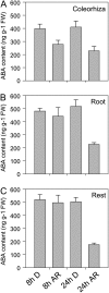
ABA quantification in barley embryo tissues. D and AR barley grains were imbibed for 8 and 24 h. Embryos were then isolated and the coleorhiza (A), root (B), and rest-of-the-embryo (C) components were dissected. ABA was isolated from the different fractions. Twenty embryos were dissected for each replicate. Three biological replicates were performed. Error bars represent se. FW, Fresh weight.
Figure 3.
Figure 3.
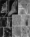
Coleorhiza anatomy in AR barley embryos during hydration. A, C, E, G, and I, Sections and images of dry embryos. B, D, F, H, and J, Images of embryos after 24 h of hydration (a 36-h-hydrated seed is shown in D). A, Confocal image of a longitudinal median section of a dry embryo showing the coleorhiza enclosing the largest embryonic root. c, Coleorhiza; ce, coleoptile; e, epiblast; m, mesocotyl; r, root; s, scutellum; sh, embryo shoot. B, Longitudinal section of an embryo following 24 h of hydration, showing elongated coleorhiza and root. The scale is the same for A and B. C, Cryo-SEM of the dry coleorhiza (indicated by the dotted line). D, Cryo-SEM of roots emerging through longitudinal cracks in the coleorhiza of a 36-h-hydrated seed. The scale is the same for C and D. E, Confocal image of coleorhiza and root cells of a dry seed. The border between the coleorhiza and the root is indicated by arrowheads. col, Coleorhiza; ep, epidermis. F, Confocal image of coleorhiza and root cells of a hydrated seed showing elongated, separated coleorhiza cells and short meristematic root cells. The scale is the same for E and F. G, Higher magnification cryo-SEM of epidermis from the coleorhiza flank of a dry seed showing small air spaces (arrowheads) between cells. H, Higher magnification cryo-SEM of a 24-h-hydrated seed showing enlarged air spaces (arrowheads) between coleorhiza epidermal cells (similar area to that shown in G). The scale is the same for G and H. I, Cryo-SEM image showing partial separation of the innermost cells of the coleorhiza in a dry seed. J, Cryo-SEM of the innermost coleorhiza cells in a hydrated seed, showing separation of the cells along their side walls, occasionally remaining attached at their end walls (arrowhead). The scale is the same for I and J.
Figure 4.
Figure 4.
Principal component (PC) analysis. This analysis was applied to the expression of all genes on the microarray over our eight samples. The numbers 8 and 18 indicate hours after imbibition. A, After-ripened; C, coleorhiza; D, dormant; R, root.
Figure 5.
Figure 5.
Global gene expression changes. Differentially expressed genes (from Supplemental Tables 2–5) at 8 or 18 h after hydration were grouped according to tissue (A and B). The same genes were also divided depending on whether they were differentially expressed in coleorhiza or in root and then grouped according to the imbibition time (C and D). The number of genes in each group is shown in parentheses.
Figure 6.
Figure 6.
Differentially expressed ABA-related genes. The expression of ABA catabolism (HvABA8′OH-1), synthesis (HvABA1, HvAAO1, and HvNCED1), signaling (HvAIP2, HvPP2C, HvLLP1, and HvLPP2), and some ABA-induced (HVA22 and Late EM abundant protein) genes is compared in the coleorhiza and root of D and AR plants. Averages of the normalized expression values of three biological replicates (Supplemental Table S1) and the se values are shown.
Figure 7.
Figure 7.
HvLPP expression during imbibition. A, Validation by real-time PCR of HvLPP1 to HvLPP3 in coleorhiza and root at 8 h after hydration. B to D, Time courses of the same genes in D and AR whole embryos at different hydration times. Averages of three biological replicates are shown. Error bars represent se.
Figure 8.
Figure 8.
ABA sensitivity assay. Time courses of germination in the dark of D and AR grains on water and on 10−4 m ABA. Germination was considered when the coleorhiza had emerged beyond the husk. Values are means of three biological replicates (20 grains were used in each replica) with their se values.
Figure 9.
Figure 9.
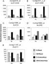
Differentially expressed light-related genes. Expression of light-related genes in D and AR coleorhiza and root is compared. The expression of two ELIP genes, HV58 and HV90, and of several light signaling components (HvHY5, HvCIP8, and PHOT1) is shown. The expression values are normalized microarray values. Data are averages of three biological replicates with se.
Figure 10.
Figure 10.
Other processes affected by after-ripening. Expression of genes related to jasmonate (HvOPR, HvAOS, HvLOX, and HvCOI1), cell wall modification (Glucan endo-1,3-β-glucosidase, XET, and β-expansin), nitrate reduction (Nitrate reductase and Nitrite reductase), and mRNA stability (Polyadenylation factor subunit and mRNA cap methyltransferase-like) is shown. The expression values are normalized microarray values. Data are averages of three biological replicates with se.
Figure 11.
Figure 11.
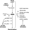
Schematic representation of the ABA regulation in barley coleorhiza by after-ripening and environmental factors. ABA has a central role blocking germination. ABA synthesis is affected by blue light (Gubler et al., 2008) and high temperature (Leymarie et al., 2008) through the expression of HvNCED1. ABA depletion is affected by after-ripening through catabolism (HvABA8′OH-1) and signaling (HvLPPs and HvAIP2). Other mechanisms affected by after-ripening identified in this paper, like cell wall modification, nitrate reduction, jasmonate metabolism, light responses, and mRNA stability, may be controlled by ABA but their action could also be ABA independent.

References

    1. Adamska I, Ohad I, Kloppstech K (1992) Synthesis of the early light-inducible protein is controlled by blue-light and related to light stress. Proc Natl Acad Sci USA 89 2610–2613 - PMC - PubMed
    1. Avery GS (1930) Comparative anatomy and morphology of embryos and seedlings of maize, oats and wheat. Bot Gaz 89 1–39
    1. Barrero JM, Jacobsen J, Gubler F (2009) Seed dormancy: approaches for finding new genes in cereals. In E-C Pua, M Davey, eds, Plant Developmental Biology: Biotechnological Perspectives. Springer, Berlin (in press)
    1. Barrero JM, Rodriguez PL, Quesada V, Piqueras P, Ponce MR, Micol JL (2006) Both abscisic acid (ABA)-dependent and ABA-independent pathways govern the induction of NCED3, AAO3 and ABA1 in response to salt stress. Plant Cell Environ 29 2000–2008 - PubMed
    1. Bartels D, Hanke C, Schneider K, Michel D, Salamini F (1992) A desiccation-related ELIP-like gene from the resurrection plant Craterostigma plantagineum is regulated by light and ABA. EMBO J 11 2771–2778 - PMC - PubMed

Publication types

MeSH terms

LinkOut - more resources