. 2009 May 1;122(Pt 9):1285-94.
doi: 10.1242/jcs.033969.

Filaggrin in the frontline: role in skin barrier function and disease

Affiliations

Filaggrin in the frontline: role in skin barrier function and disease

Aileen Sandilands et al. J Cell Sci. .

Abstract

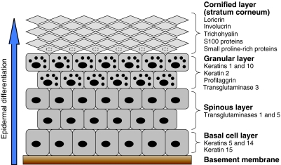
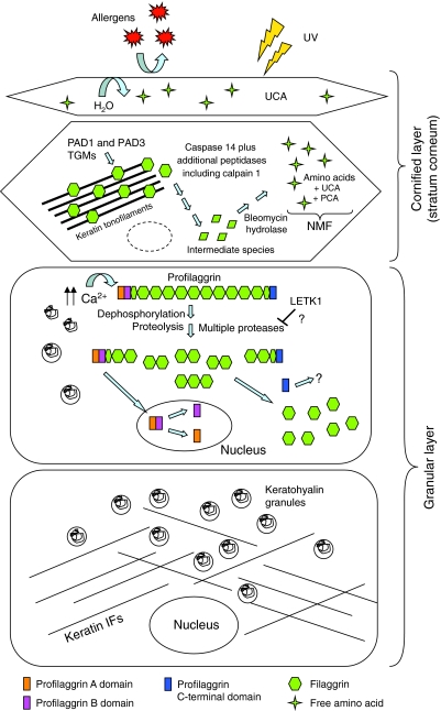
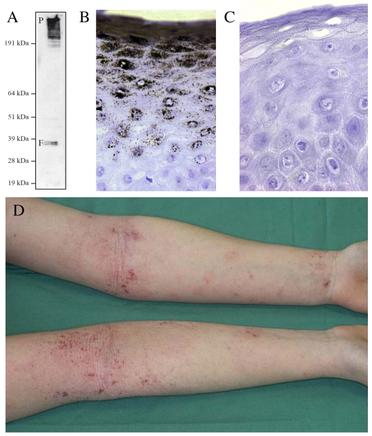
Recently, loss-of-function mutations in FLG, the human gene encoding profilaggrin and filaggrin, have been identified as the cause of the common skin condition ichthyosis vulgaris (which is characterised by dry, scaly skin). These mutations, which are carried by up to 10% of people, also represent a strong genetic predisposing factor for atopic eczema, asthma and allergies. Profilaggrin is the major component of the keratohyalin granules within epidermal granular cells. During epidermal terminal differentiation, the approximately 400 kDa profilaggrin polyprotein is dephosphorylated and rapidly cleaved by serine proteases to form monomeric filaggrin (37 kDa), which binds to and condenses the keratin cytoskeleton and thereby contributes to the cell compaction process that is required for squame biogenesis. Within the squames, filaggrin is citrullinated, which promotes its unfolding and further degradation into hygroscopic amino acids, which constitute one element of natural moisturising factor. Loss of profilaggrin or filaggrin leads to a poorly formed stratum corneum (ichthyosis), which is also prone to water loss (xerosis). Recent human genetic studies strongly suggest that perturbation of skin barrier function as a result of reduction or complete loss of filaggrin expression leads to enhanced percutaneous transfer of allergens. Filaggrin is therefore in the frontline of defence, and protects the body from the entry of foreign environmental substances that can otherwise trigger aberrant immune responses.


Figures

Fig. 1.
Fig. 1.
Epidermal differentiation. The epidermis is the outermost layer of the skin and is separated from the underlying dermis by the basement membrane. Keratinocytes, which compose the epidermis, proliferate within the basal cell layer. As differentiation proceeds, keratinocytes progress upwards through the different epidermal layers (the spinous layer, granular layer and cornified layer or stratum corneum), becoming anucleated and increasingly compacted in size, before being eventually lost from the skin surface by desquamation (shedding of the outer layers of skin). Each stage of epidermal differentiation is characterised by the expression of specific proteins, and examples of these are listed on the figure. The smaller black dots in the cells of the granular layer represent keratohyalin granules.
Fig. 2.
Fig. 2.
(A) Profilaggrin and filaggrin gene structure. The FLG gene, which is located within the epidermal differentiation complex on chromosome 1q21, spans ∼25 kb of DNA and comprises three exons and two introns. The majority of the profilaggrin protein is encoded by exon 3. (B) Profilaggrin protein structure. Profilaggrin is expressed as a polyprotein that contains a variable number (10-12) of tandemly arranged, near-identical full-length filaggrin repeats, which are flanked on either side by partial, imperfect filaggrin repeats. Each repeat is 324 amino acids long and is separated from other repeats by a short linker. Within the N-terminal domain, the A domain contains two Ca2+-binding motifs that have similarity to the EF-hands of the S100 protein family. The B domain of the N-terminal region and the C-terminal domain are also shown. (C) Profilaggrin linker region. A comparison of the amino acid sequences of the proposed profilaggrin linker regions in mouse, rat and human. The linker region of human profilaggrin is shorter than those of mouse and rat, and differs significantly in its amino acid composition. Within human profilaggrin, the sequence of the linker region between the individual filaggrin repeats is highly conserved. Asterisks denote amino acid residues that are phosphorylated in mouse and rat (Resing et al., 1985; Resing et al., 1995a). Shaded residues indicate those that are conserved among two or three of the sequences.
Fig. 3.
Fig. 3.
Phosphorylation of profilaggrin. (A) Profilaggrin phosphorylation sites. Clustering of phosphorylated residues (open arrowheads) within the filaggrin repeats of rat profilaggrin. The size of the arrowhead reflects the number of phosphorylated residues. The linker sequence between filaggrin repeats contains two phosphorylated residues. A cluster of seven phosphorylated residues is found upstream of the linker and a comparison of the amino acid sequences of rat, mouse and human in this cluster is shown above. Asterisks denote amino acid residues that are phosphorylated in rat (Resing et al., 1995a). (B) Putative protein-kinase target sites of human profilaggrin. Protein kinases that might target profilaggrin were identified using motif-prediction software (http://scansite.mit.edu/) and from known protein-kinase consensus sequences. The different font sizes reflect the degree of similarity between the potential recognition sites in profilaggrin and the consensus recognition sequence of the kinase - the smaller the font size, the lower the degree of similarity. PKB/Akt, protein kinase B; CDK5, cyclin-dependent kinase 5; Y-kinase, tyrosine kinase; DNA-PK, DNA-dependent protein kinase; GSK3, glycogen synthase kinase 3; AGC/CAMK kinases, members of the AGC and CAMK kinase families.
Fig. 4.
Fig. 4.
Profilaggrin processing during terminal differentiation of the epidermis. Within the granular layer, profilaggrin is stored in an inactive and insoluble form within keratohyalin granules. In response to an increase in Ca2+ levels, the keratohyalin granules degranulate and profilaggrin is dephosphorylated and proteolysed in a multistep process into free filaggrin monomers by a variety of proteases including matriptase, prostasin and probably kallikrein 5 (see text for details). Following cleavage from the filaggrin monomers the N-terminal head domain undergoes nuclear translocation and further degradation into the A and B domains. In the cornified layer (stratum corneum), the released filaggrin monomers bind directly to keratin filaments, causing their collapse into thickened and aggregated keratin filaments, which has the effect of condensing the keratinocyte cytoskeleton. Condensation of the cytoskeleton is followed by crosslinking with transglutaminases (TGMs) and modification by peptidylarginine deiminases (PADs) to form an insoluble keratin matrix. Together with lipids and other cornified-layer proteins, this ultimately forms the so-called `skin barrier', which prevents water loss through the skin as well as the unwanted entry of molecules such as allergens. Filaggrin undergoes subsequent degradation by a variety of proteases, including caspase 14, into free amino acids and derivatives such as urocanic acid (UCA) and pyrrolidone carboxylic acid (PCA) - these are collectively referred to as natural moisturising factor (NMF), which contributes to skin hydration and possibly to UV protection.
Fig. 5.
Fig. 5.
Filaggrin in human disease. (A) Post-translational processing of profilaggrin to filaggrin. Immunoblot of high-salt protein extract of human epidermis probed with monoclonal antibody 15C10 (Novacastra, Newcastle upon Tyne, UK), which recognises an epitope within the filaggrin repeat domain and therefore detects both profilaggrin (P, upper band, ∼400 kDa) and processed filaggrin (F, lower doublet, ∼37 kDa). (B) Immunohistochemical straining of human epidermis with 15C10, showing the great abundance of profilaggrin and/or filaggrin in the upper granular layers of the epidermis. (C) Immunohistochemical staining of epidermis derived from an individual homozygous for nonsense mutation R501X in the first filaggrin repeat (compare with B). Filaggrin is completely absent in this individual, who has severe ichthyosis vulgaris, atopic eczema and other allergies. A recent population study in the north of England showed that approximately 1 in 90 children have an equivalent filaggrin-null genotype (Brown et al., 2008). (D) A patient with atopic eczema showing the flexural (referring to skin folds, such as the inner surface of elbows) inflammation that is a classic clinical hallmark of this common, complex trait. Filaggrin-null mutations represent a major genetic risk factor for atopic eczema.

Similar articles

Cited by

References

    1. Brown, S. J., Relton, C. L., Liao, H., Zhao, Y., Sandilands, A., Wilson, I. J., Burn, J., Reynolds, N. J., McLean, W. H. I. and Cordell, H. J. (2008). Filaggrin null mutations and childhood atopic eczema: a population-based case-control study. J. Allergy Clin. Immunol. 121, 940-946.e3. - PMC - PubMed
    1. Candi, E., Rufini, A., Terrinoni, A., Giamboi-Miraglia, A., Lena, A. M., Mantovani, R., Knight, R. and Melino, G. (2007). DeltaNp63 regulates thymic development through enhanced expression of FgfR2 and Jag2. Proc. Natl. Acad. Sci. USA 104, 11999-12004. - PMC - PubMed
    1. Candi, E., Cipollone, R., Rivetti di Val Cervo, P., Gonfloni, S., Melino, G. and Knight, R. (2008). p63 in epithelial development. Cell Mol. Life Sci. 65, 3126-3133. - PMC - PubMed
    1. Chavanas, S., Bodemer, C., Rochat, A., Hamel-Teillac, D., Ali, M., Irvine, A. D., Bonafe, J. L., Wilkinson, J., Taieb, A., Barrandon, Y. et al. (2000). Mutations in SPINK5, encoding a serine protease inhibitor, cause Netherton syndrome. Nat. Genet. 25, 141-142. - PubMed
    1. Chen, H., Ho, J. C., Sandilands, A., Chan, Y. C., Giam, Y. C., Evans, A. T., Lane, E. B. and McLean, W. H. I. (2008). Unique and recurrent mutations in the filaggrin gene in Singaporean Chinese patients with ichthyosis vulgaris. J. Invest. Dermatol. 128, 1669-1675. - PubMed

Publication types

MeSH terms