Comparative Study
. 2009 Apr 22;29(16):5100-7.
doi: 10.1523/JNEUROSCI.4149-08.2009.

Role of protein kinase C in the induction and maintenance of serotonin-dependent enhancement of the glutamate response in isolated siphon motor neurons of Aplysia californica

Affiliations
Comparative Study

Role of protein kinase C in the induction and maintenance of serotonin-dependent enhancement of the glutamate response in isolated siphon motor neurons of Aplysia californica

Greg Villareal et al. J Neurosci. .

Abstract

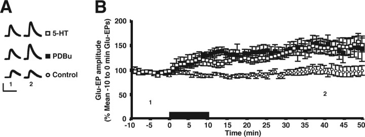
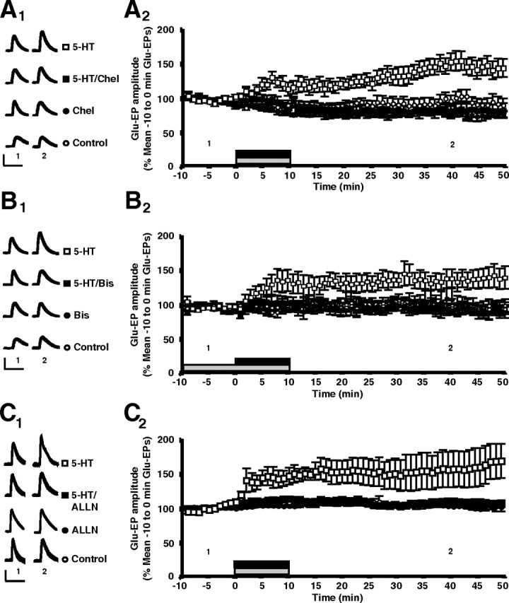
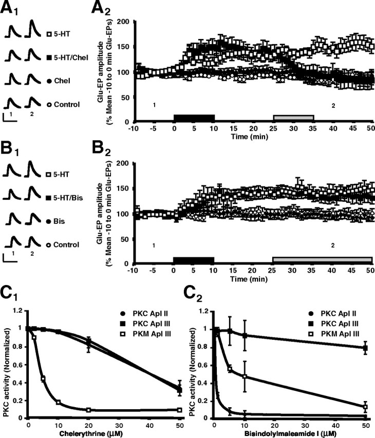
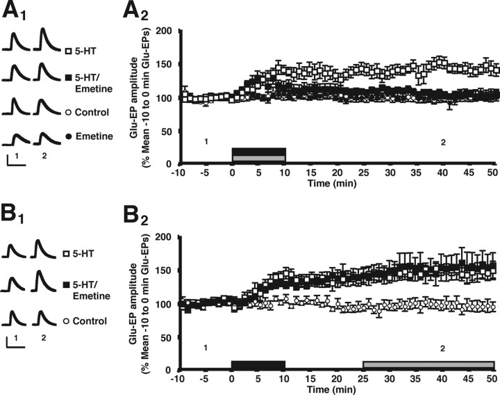
Serotonin (5-HT) mediates learning-related facilitation of sensorimotor synapses in Aplysia californica. Under some circumstances 5-HT-dependent facilitation requires the activity of protein kinase C (PKC). One critical site of PKC's contribution to 5-HT-dependent synaptic facilitation is the presynaptic sensory neuron. Here, we provide evidence that postsynaptic PKC also contributes to synaptic facilitation. We investigated the contribution of PKC to enhancement of the glutamate-evoked potential (Glu-EP) in isolated siphon motor neurons in cell culture. A 10 min application of either 5-HT or phorbol ester, which activates PKC, produced persistent (> 50 min) enhancement of the Glu-EP. Chelerythrine and bisindolylmaleimide-1 (Bis), two inhibitors of PKC, both blocked the induction of 5-HT-dependent enhancement. An inhibitor of calpain, a calcium-dependent protease, also blocked 5-HT's effect. Interestingly, whereas chelerythrine blocked maintenance of the enhancement, Bis did not. Because Bis has greater selectivity for conventional and novel isoforms of PKC than for atypical isoforms, this result implicates an atypical isoform in the maintenance of 5-HT's effect. Although induction of enhancement of the Glu-EP requires protein synthesis (Villareal et al., 2007), we found that maintenance of the enhancement does not. Maintenance of 5-HT-dependent enhancement appears to be mediated by a PKM-type fragment generated by calpain-dependent proteolysis of atypical PKC. Together, our results suggest that 5-HT treatment triggers two phases of PKC activity within the motor neuron, an early phase that may involve conventional, novel or atypical isoforms of PKC, and a later phase that selectively involves an atypical isoform.


Figures

Figure 1.
Figure 1.
Both 5-HT and phorbol ester enhance the glutamate response in Aplysia motor neurons. A, Comparison of the effects of 5-HT and PDBu on the glutamate-evoked potentials (Glu-EPs). Each pair of traces represents responses from a single experiment, which lasted 1 h. The drug, either 5-HT or PDBu, was applied for 10 min. In the control experiments, the vehicle alone was applied. Traces marked “1” represent Glu-EPs evoked 5 min before the onset of drug treatment (or 5 min after the start of testing); those marked “2” represent Glu-EPs evoked 30 min after washout of the drug (or 50 min after the start of testing). Calibration: 10 mV, 500 ms. B, Summary of the enhancement of the Glu-EP produced by 5-HT or PDBu. The motor neurons were stimulated with a glutamate test pulse once per 10 s; each symbol represents the mean normalized amplitude of six consecutive Glu-EPs. After 10 min of glutamate stimulation to establish a baseline response, motor neurons received either 5-HT (open squares), PDBu (filled squares), or vehicle alone (control group, open circles). Drug treatment period is indicated by the black bar. The numbers below the data indicate the trials during which the sample Glu-EPs shown in A were recorded. Error bars represent ± SEM.
Figure 2.
Figure 2.
Inhibition of either PKC or calpain blocks the induction of 5-HT-dependent enhancement of the glutamate response. A1, Effect of the PKC inhibitor chelerythrine on the induction of enhancement of the Glu-EP. The figure presents sample traces from a single experiment for each experimental group. Note that in the control experiments, no drug was applied. Traces marked “1” represent Glu-EPs evoked 5 min before the onset of the 10 min period of drug treatment (or 5 min after the start of testing); those marked “2” represent Glu-EPs evoked 30 min after drug washout (or 50 min after the start of testing). Calibration: A1, B1, C1, 10 mV, 500 ms. A2, Summary of the experiments showing the effect of chelerythrine on induction. Motor neurons received either 5-HT alone (open squares), 5-HT combined with chelerythrine (filled squares), chelerythrine alone (Chel; filled circles), or perfusion medium alone (controls; open circles). The black bar indicates the period of 5-HT treatment; the gray bar indicates the period of chelerythrine treatment. The numbers below the data indicate the trials during which the sample Glu-EPs shown in A1 were recorded. Error bars represent ± SEM. B1, Effect of another PKC inhibitor, bisindolylmaleimide-1 (Bis), on the induction of enhancement of the glutamate response. Shown are sample traces from a single experiment for each experimental group. In the control experiments only the vehicle was applied. The Bis was applied for 20 min, and was present for 10 min before the onset of the 10 min period of 5-HT or treatment, which occurred 10 min after the start of testing. Traces marked “1” represent Glu-EPs evoked 5 min before the onset of 5-HT or vehicle (or 5 min after the start of testing); those marked “2” represent Glu-EPs evoked 30 min after washout of the drug or vehicle (or 50 min after the start of testing). B2, Summary of the experiments showing block of the induction of 5-HT-dependent enhancement by Bis. Motor neurons were treated with 5-HT (open squares), 5-HT and Bis (filled squares), Bis alone (filled circles), or vehicle alone (control; open circles). The black bar indicates the 5-HT treatment period, whereas the gray bar indicates the Bis treatment period. The numbers below the data indicate the trials at which the sample Glu-EPs shown in B1 were recorded. Error bars represent ± SEM. C1, Effect of the calpain inhibitor ALLN on the induction of enhancement. The figure shows sample traces from a single experiment for each experimental group. In the control experiments no drug was applied. Traces marked “1” represent Glu-EPs evoked 5 min before the onset of drug treatment (or 5 min after the start of testing); those marked “2” represent Glu-EPs evoked 30 min after drug washout (or 50 min after the start of testing). C2, Summary of the experiments showing the block of enhancement of the Glu-EP by inhibition of calpain activity. Motor neurons received 5-HT (open squares), 5-HT plus ALLN (filled squares), ALLN alone (filled circles), or perfusion medium alone (control; open circles). The black bar indicates the 5-HT treatment period, whereas the gray bar indicates the ALLN treatment period. The numbers below the data indicate the trials at which the sample Glu-EPs shown in C1 were recorded. Error bars represent ± SEM.
Figure 3.
Figure 3.
PKM Apl III mediates maintenance of 5-HT-dependent enhancement. A1, Effect of chelerythrine on the maintenance of the enhancement. The figure presents sample traces from a single experiment for each experimental group. No drug was applied in the control experiments. Traces marked “1” represent Glu-EPs evoked 5 min before the onset of 5-HT treatment (or 5 min after the start of testing); those marked “2” represent Glu-EPs evoked 30 min after washout of 5-HT (or 50 min after the start of testing). In the experiments involving chelerythrine, the inhibitor was applied for 10 min starting 15 min after washout of 5-HT, or during the equivalent time period in the chelerythrine-alone experiments. Calibration: A1, B1, 10 mV, 500 ms. A2, Summary of the experiments showing that chelerythrine disrupts the maintenance of enhancement. Motor neurons were stimulated with 5-HT alone (open squares), 5-HT followed by chelerythrine (25–35 min trials; filled squares), chelerythrine alone (filled circles), or perfusion medium alone (control; open circles). The black bar indicates the period of 5-HT treatment, whereas the gray bar indicates the chelerythrine treatment period. Notice that the enhancement did not reappear after washout of chelerythrine in the 5-HT/Chel experiments. The numbers below the data indicate the trials at which the sample Glu-EPs shown in A1 were recorded. Error bars represent ± SEM. B1, Effect of Bis on the maintenance of 5-HT-dependent enhancement of the Glu-EP. The figure presents sample traces from a single experiment for each experimental group. Traces marked “1” represent Glu-EPs evoked 5 min before the onset of 5-HT treatment (or 5 min after the start of testing); those marked “2” represent Glu-EPs evoked 30 min after washout of 5-HT (or 50 min after the start of testing). In the experiments involving Bis, the inhibitor was applied for 25 min starting 15 min after washout of 5-HT, or during the equivalent time period in the Bis-alone experiments. In the control experiments the vehicle was applied during the same time that Bis was applied in the 5-HT/Bis and Bis-alone experiments. B2, Summary of the experiments showing the lack of an effect of Bis on maintenance of the enhancement. Cultures were exposed to 5-HT (open squares), 5-HT followed by Bis (25–50 min trials; filled squares), Bis alone (filled circles), or vehicle alone (control; open circles). The black bar indicates the 5-HT treatment period, whereas the gray bar indicates the Bis treatment period. The numbers below the data indicate the trials at which the sample Glu-EPs shown in B1 were recorded. Error bars represent ± SEM. C1, Inhibition of PKC Apl II (filled circles), PKC Apl III (filled squares), and PKM Apl III (open squares) by chelerythrine. C2, Inhibition of PKC Apl II (filled circles), PKC Apl III (filled squares), and PKM Apl III (open squares) by Bis. Error bars are ± SEM; n > 3 for each concentration.
Figure 4.
Figure 4.
Although induction of the enhancement of the glutamate response requires protein synthesis, maintenance of the enhancement does not depend on ongoing protein synthesis. A1, Effect of emetine treatment on the induction of 5-HT-dependent enhancement of the Glu-EP. The figure presents sample traces from a single experiment for each experimental group. No drug was applied in the control experiments. Traces marked “1” represent Glu-EPs evoked 5 min before the onset of drug treatment (or 5 min after the start of testing); those marked “2” represent Glu-EPs evoked 30 min after drug washout (or 50 min after the start of testing). Calibration: A1, B1, 10 mV, 500 ms. A2, Summary of experiments showing that inhibition of protein synthesis blocks the induction of the enhancement of the Glu-EP. Motor neurons received either 5-HT alone (open squares), 5-HT and emetine (filled squares), emetine alone (filled circles), or perfusion medium alone (control; open circles). The overall mean Glu-EP in the 5-HT-alone group (131 ± 1%, n = 8) was significantly greater than those in groups treated with 5-HT and emetine (109 ± 1%, n = 7) and emetine alone (102 ± 1%, n = 6), as well as in the control group (101 ± 1%, n = 6) (p < 0.05 for each comparison). The black bar indicates the 5-HT treatment, and the gray bar the emetine treatment. The numbers below the data indicate the trials at which the sample Glu-EPs shown in A1 were recorded. Error bars represent ± SEM. From Villareal et al. (2007). See that study for additional details. B1, Effect of emetine treatment on the maintenance of 5-HT-dependent enhancement of the Glu-EP. The figure presents sample traces from a single experiment for each experimental group. No drug was applied in the control experiments. Traces marked “1” represent Glu-EPs evoked 5 min before the onset of 5-HT treatment (or 5 min after the start of testing); those marked “2” represent Glu-EPs evoked 30 min after washout of 5-HT (or 50 min after the start of testing). B2, Summary of experiments showing that protein synthesis is not required for the maintenance of 5-HT-dependent enhancement. Motor neurons received either 5-HT alone (open squares), 5-HT followed by emetine (filled squares), or perfusion medium alone (control, open circles). In the experiments involving emetine, the inhibitor was applied for 25 min starting 15 min after washout of 5-HT. The black bar indicates the 5-HT treatment period, and the gray bar indicates the emetine treatment period. The numbers below the data indicate the trials at which the sample Glu-EPs shown in B1 were recorded. Error bars represent ± SEM.

Similar articles

Cited by

References

    1. Bougie J, Lim T, Ferraro G, Manjunath V, Scott D, Sossin WS. Cloning and characterization of protein kinase C (PKC) Apl III, a homologue of atypical PKCs in Aplysia . Soc Neurosci Abstr. 2006;32:669–10.
    1. Bougie JK, Lim T, Manjunath V, Farah-Abi C, Nagakura I, Sossin WS. The role of atypical protein kinase C (PKC) zeta in synaptic plasticity in Aplysia . Soc Neurosci Abstr. 2007;33:208–5.
    1. Bougie JK, Lim T, Farah CA, Manjunath V, Nagakura I, Ferraro GB, Sossin WS. The atypical protein kinase C in Aplysia can form a PKM by cleavage. J Neurochem. 2009 doi: 10.1111/j.1471-4159.2009.06045.x. - DOI - PMC - PubMed
    1. Braha O, Dale N, Hochner B, Klein M, Abrams TW, Kandel ER. Second messengers involved in the two processes of presynaptic facilitation that contribute to sensitization and dishabituation in Aplysia sensory neurons. Proc Natl Acad Sci U S A. 1990;87:2040–2044. - PMC - PubMed
    1. Brunelli M, Castellucci V, Kandel ER. Synaptic facilitation and behavioral sensitization in Aplysia: possible role of serotonin and cyclic AMP. Science. 1976;194:1178–1181. - PubMed

Publication types

MeSH terms

LinkOut - more resources