Comparative Study
. 2009 Apr 22;29(16):5127-34.
doi: 10.1523/JNEUROSCI.0859-09.2009.

NAD+ activates KNa channels in dorsal root ganglion neurons

Affiliations
Comparative Study

NAD+ activates KNa channels in dorsal root ganglion neurons

Thomas J Tamsett et al. J Neurosci. .

Abstract

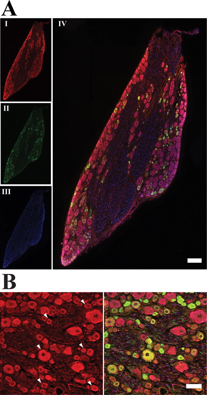
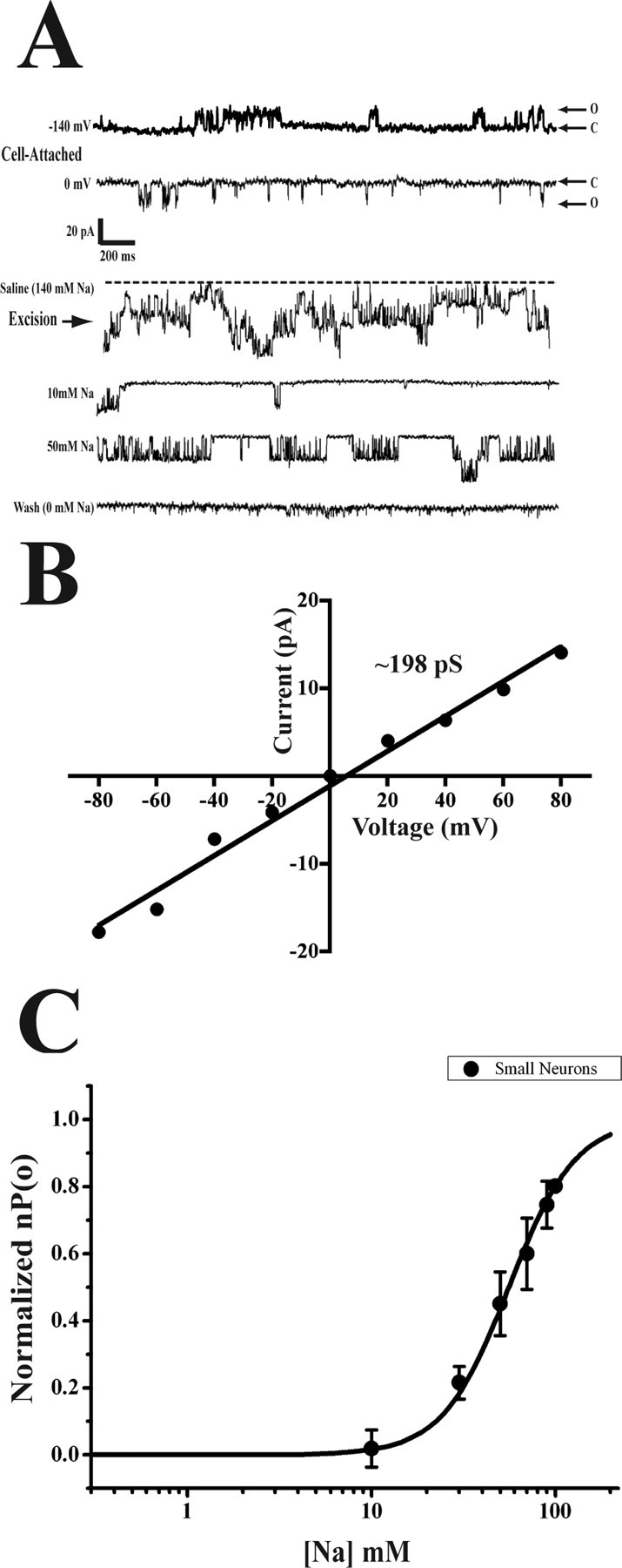
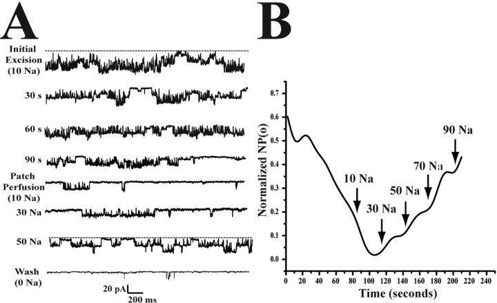
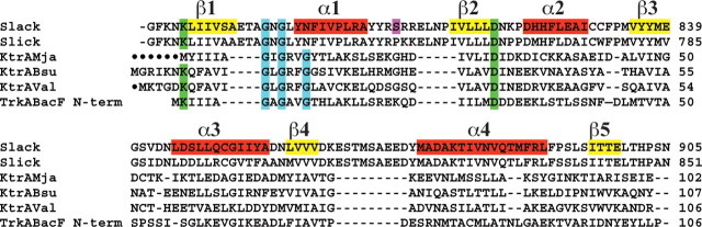
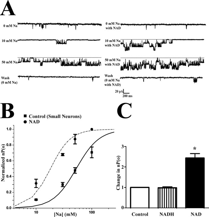
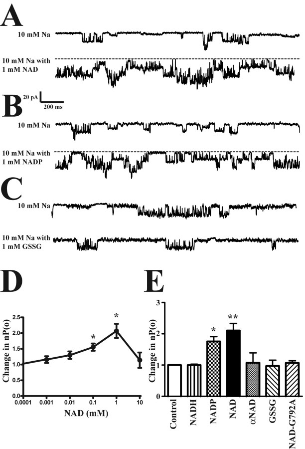
Although sodium-activated potassium channels (KNa) have been suggested to shape various firing patterns in neurons, including action potential repolarization, their requirement for high concentrations of Na+ to gate conflicts with this view. We characterized KNa channels in adult rat dorsal root ganglion (DRG) neurons. Using immunohistochemistry, we found ubiquitous expression of the Slack KNa channel subunit in small-, medium-, and large-diameter DRG neurons. Basal KNa channel activity could be recorded from cell-attached patches of acutely dissociated neurons bathed in physiological saline, and yet in excised inside-out membrane patches, the Na+ EC50 for KNa channels was typically high, approximately 50 mM. In some cases, however, KNa channel activity remained considerable after initial patch excision but decreased rapidly over time. Channel activity was restored in patches with high Na+. The channel rundown after initial excision suggested that modulation of channels might be occurring through a diffusible cytoplasmic factor. Sequence analysis indicated that the Slack channel contains a putative nicotinamide adenine dinucleotide (NAD+)-binding site; accordingly, we examined the modulation of native KNa and Slack channels by NAD+. In inside-out-excised neuronal patch recordings, we found a decrease in the Na+ EC50 for KNa channels from approximately 50 to approximately 20 mM when NAD+ was included in the perfusate. NAD+ also potentiated recombinant Slack channel activity. NAD+ modulation may allow KNa channels to operate under physiologically relevant levels of intracellular Na+ and hence provides an explanation as to how KNa channel can control normal neuronal excitability.


Figures

Figure 1.
Figure 1.
Slack is ubiquitously expressed in adult rat DRG. A, Whole DRG staining. Multiple lower magnification single confocal images were compiled together into a single image representing an entire rat DRG. I, Slack; II, CGRP; III, neurofilament; IV, overlay of I–III. More than 90% DRG neurons had Slack staining. B, Higher magnification. Cell bodies and axons exhibited Slack labeling. Arrowheads indicate cell surface localization. Scale bars: A, 200 μm; B, 50 μm.
Figure 2.
Figure 2.
KNa channels recorded from adult small DRG neurons. A, Large conductance “KNa-like” channels recorded in cell-attached patches on small DRG neurons bathed in physiological saline (140 mm Na+ and 3 mm K+). Pipette solution contained 130 mm K+ and 1 mm TEA. Voltage indicated is the applied pipette potential. Excision into high external Na+ confirmed KNa channels. Patches were subsequently perfused with varying concentrations of Na+ to confirm Na+ dependence. Channels here were recorded at holding potential of −80 mV. B, Current–voltage relationship in symmetrical K+ revealed a large conductance channel. C, Na+ dose–response in small neurons (n = 6). EC50, 53 mm; Hill coefficient, 2.4.
Figure 3.
Figure 3.
Rundown of KNa channels after patch excision. A, Representative traces of high basal KNa channel activity. Rundown occurred in three of six patches from small DRG neurons. B, NP(o) values were calculated for three patches every 2 s after patch excision in inside-out configuration. Channels were recorded at −80 mV. The nP(o)s were averaged after every 10 s, normalized to highest nP(o) in each neuron, and the averages for each time point were taken. Arrows indicate approximate commencement of perfusion of patches at each concentration of Na+ (all values in millimolars). A Savitzky–Golay smoothing curve was used to filter data.
Figure 4.
Figure 4.
Slack and Slick contain putative NAD+-binding sites. NAD+-binding sequence in Slack, Slick, and known NAD+-dependent K+ transporters from Methanocaldococcus jannaschii (Mja), Bacillus subtilis (Bsu), Vibro alginolyticus (Val), and Bacteroides fragilis (BacF). Yellow highlights indicate β sheets, whereas red highlights indicate α helices. The fingerprint region consists of a βαβαβ structure. The first green highlights indicated the conserved positive charge in beginning of the first β sheet in the fingerprint region. The second green highlights indicate the conserved negatively charged residue at the end of the second β sheet in the fingerprint region. The blue highlights are the phosphate-binding consensus sequence “GXGXXG” connecting the first β sheet to the first α helix. The purple highlights a consensus protein kinase C phosphorylation site present in Slack but not in Slick.
Figure 5.
Figure 5.
NAD+ modulates KNa channels in small DRG neurons. A, Representative trace of channels recorded in an inside-out-excised patch from a small DRG neuron with X mm Na+ (control) and with X mm Na+ and 1 mm NAD+. Voltage held at −80 mV. B, Na+ dose–response relationship of control and NAD+ perfusions in small adult rat DRG neurons. NP(o) values were calculated from 30 sweeps of 2400 ms time samples at each dose of Na+ and Na+/NAD+ solution. Each point represents an n = 6. Error bars represent SEM. EC50 was determined to be 50 mm for Na+ and 17 mm for Na+/NAD+ solutions in small adult DRG neurons with a Hill coefficient of 2.4. C, NADH does not modulate KNa channel activity in DRG neurons. Na+ (30 mm; control) was perfused onto inside-out-excised patches and compared with 30 mm Na+ + 1 mm NADH and 30 mm + 1 mm NAD+. Voltage was held at −80 mV. NP(o) values were calculated from 30 sweeps of 2400 ms time samples (n = 5; *p < 0.01). Error bars represent SEM.
Figure 6.
Figure 6.
NAD+ and NADP+ modulate Slack channels stably expressed in HEK-293T. A, Representative traces of NAD+-activating recombinant Slack channels. B, Representative traces of NADP+-activating recombinant Slack channels. C, Representative traces of GSSG failing to activate recombinant Slack channels. All voltages held at −80 mV. Excised patch recordings were performed from a Slack-stable-transfected HEK-293 cell line. D, NAD+ dose–response relationship on Slack channels. Each perfusion contained X mm NAD+, 10 mm Na+, 40 mm K+, 90 mm NMG. *p < 0.01, statistical significance compared with control. E, Summary of the effects of NADH, NADP+, NAD+, αNAD+, the oxidizing agent, GSSG, on Slack channels and NAD+ on a Slack G792A mutant channel versus the control. The Slack mutant was generated through by site-directed mutagenesis of second glycine in NAD+ fingerprint region. All solutions contained 1 mm of either NAD+, NADH or NADP+, αNAD+, GSSG, and 10 mm Na+, 40 mm K+, 90 mm NMG. *p < 0.005 and **p < 0.001, significant difference versus control. Error bars represent SEM.

References

    1. Amir R, Michaelis M, Devor M. Membrane potential oscillations in dorsal root ganglion neurons: role in normal electrogenesis and neuropathic pain. J Neurosci. 1999;19:8589–8596. - PMC - PubMed
    1. Bellamacina CR. The nicotinamide dinucleotide binding motif: a comparison of nucleotide binding proteins. FASEB J. 1996;10:1257–1269. - PubMed
    1. Bhattacharjee A, Kaczmarek LK. For K+ channels, Na+ is the new Ca2+ Trends Neurosci. 2005;28:422–428. - PubMed
    1. Bhattacharjee A, Gan L, Kaczmarek LK. Localization of the Slack potassium channel in the rat central nervous system. J Comp Neurol. 2002;454:241–254. - PubMed
    1. Bhattacharjee A, Joiner WJ, Wu M, Yang Y, Sigworth FJ, Kaczmarek LK. Slick (Slo2.1), a rapidly-gating sodium-activated potassium channel inhibited by ATP. J Neurosci. 2003;23:11681–11691. - PMC - PubMed

Publication types

MeSH terms

LinkOut - more resources