Review
. 2009 May 1;6(3):249-55.
doi: 10.1513/pats.200806-054RM.

Nuclear factor kappaB, airway epithelium, and asthma: avenues for redox control

Affiliations
Review

Nuclear factor kappaB, airway epithelium, and asthma: avenues for redox control

Yvonne M W Janssen-Heininger et al. Proc Am Thorac Soc. .

Abstract

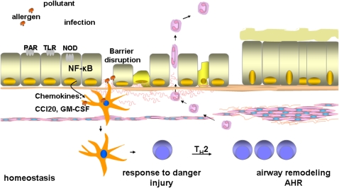
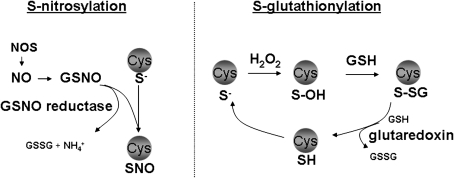
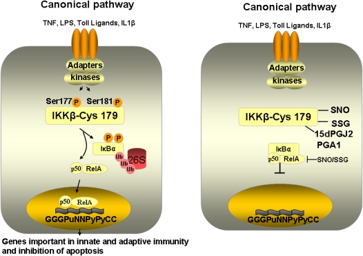
A wealth of recent studies points to the importance of airway epithelial cells in the orchestration of inflammatory responses in the allergic inflamed lung. Studies also point to a role of oxidative stress in the pathophysiology of chronic inflammatory diseases. This article provides a perspective on the significance of airway epithelial cells in allergic inflammation, and reviews the relevance of the transcription factor, nuclear factor kappaB, herein. We also provide the reader with a perspective on the role that oxidants can play in lung homeostasis, and address the concept of "redox biology." In addition, we review recent evidence that highlights potential inhibitory roles of oxidants on nuclear factor kappaB activation and inflammation, and discuss recent assays that have become available to probe the functional roles of oxidants in lung biology.


Figures

<b>Figure 1.</b>
Figure 1.
Schematic presentation of the functional role of airway epithelium in the pathogenesis of asthma. Epithelial cells, which are positioned at the interface with the environment, can sense and respond to inhaled antigens, allergens, pollutants, proteases, microbes, through the activation of Toll-like receptors (TLR), Nod-like receptors (NOD), and protease activated receptors (PAR), and others, leading to the activation of nuclear factor κB (NF-κB). Activation of NF-κB leads to the transcriptional activation of many proinflammatory genes that include cytokine and chemokines, granulocyte macrophage–colony-stimulating factor (GM-CSF), and CC chemokine ligand 20 (CCL20). Disruption of the epithelial barrier as a result of inhaled pollutants, or proteases present in allergens, results in enhanced access of antigens to dendritic cells (DCs). The increased recruitment and activation of DCs through the actions of CCL20 and GM-CSF, and enhanced accessibility of antigen result in maturation of DCs, and their polarization, leading to subsequent Th2 immune responses, critical to airway remodeling.
<b>Figure 2.</b>
Figure 2.
Overview of NF-κB activation pathways. In the canonical NF-κB activation pathway, which is stimulated by ligands such as tumor necrosis factor (TNF)-α, lipopolysaccharide (LPS), and interleukin (IL)-1β, IKKβ is responsible for the phosphorylation of IκBα, leading to its subsequent ubiquitination and degradation through the proteasome pathway. This allows for translocation of RelA/p50 to the nucleus, and the subsequent activation of transcription of genes important in innate immunity and protection from apoptosis. In the noncanonical (alternative) NF-κB activation pathway, which is activated through ligands such as lymphotoxin β (LTB), B cell–activating factor (BAFF), and CD40 ligand (CD40L), among others, NF-κB–inducing kinase (NIK)-dependent activation of IKKα leads to phoshorylation of p100, subsequent ubiquitination, and proteolytic processing to p52. This results in nuclear translocation of p52/RelB complexes, and activation of distinct transcriptional programs. For illustrative purposes, this is a simplified overview. Additional phosphorylation events are induced by IKK proteins, notably the phosphorylation of histone H3, CREB-binding protein, and silencing mediator of retinoic acid and thyroid hormone receptor by IKKα. Additional post-translational modifications of NF-κB members that include phosphorylation, ubiquitination and acetylation, function of transcriptional co-activators and repressors, and chromatin remodeling events are collectively important to shape the nature, strength, and duration of the NF-κB transcriptional response. While the canonical and noncanonical pathways are drawn separately for illustrative purposes, we point out that these pathways do not act independently. Both cooperative and inhibitory roles in reciprocal regulation have been reported, indicated by arrows (see References and for recent reviews).
<b>Figure 3.</b>
Figure 3.
Overview of the biochemical events that control protein S-nitrosylation (SNO) and S-glutathionylation. Nitric oxide synthases (NOS) produce nitric oxide (NO), which can become converted to S-nitrosothiols, including S-nitrosoglutathione (GSNO). S-nitrosothiols can cause protein S-nitrosylation, which exerts important regulatory functions in target proteins. The enzyme, GSNO reductase, metabolizes GSNO and produces ammonia (NH4+) and oxidized glutathione (GSSG), and indirectly controls protein S-nitrosylation. Hydrogen peroxide (H2O2) can oxidize protein cysteines that are in the thiolate state (S), causing the formation of unstable sulfenic acid (S-OH) intermediates, which are targets for S-glutathionylation (P-SSG). In physiologic settings, mammalian glutaredoxins serve to reduce the S-glutathionylated proteins, restoring the sulfhydryl (SH) group, thus regulating the extent of PSSG. For detailed information see Reference .
<b>Figure 4.</b>
Figure 4.
Overview of activation of the canonical (classical) NF-κB pathway, and visualization of the inhibitory effect of cysteine oxidation of IKKβ at cysteine 179 on activation of the NF-κB pathway. We have also highlighted that the same cysteine is targeted by the antiinfammatory prostaglandins, PGA1 and 15dPGJ2. 15dPGJ2 = 15-deoxy-Δ12–14-prostaglandin J2; PGA1 = prostaglandin A1; SNO = S-nitrosylation; SSG = S-glutathionylation. Inhibition of RelA and p50 after S-nitrosylation and S-glutathionylation is also highlighted.
<b>Figure 5.</b>
Figure 5.
Detection of protein S-nitrosylation (PSNO) and protein S-glutathionylation (PSSG) in intact C10 lung epithelial cells. Control cells were fixed and subjected to thiol blocking, ascorbate dependent decomposition of S-nitrosylated proteins, and subsequent biotin labeling of newly generated reduced sulfhydryls using a biotin-conjugated probe, incubation with streptavidin-conjugated fluorophore, and detection via confocal laser scanning flow cytometry. S-glutathionylated proteins were visualized after glutaredoxin-catalyzed reduction of S-glutathionylated proteins. Details of these procedures were reviewed elsewhere (29).

References

    1. Bousquet J, Jeffery PK, Busse WW, Johnson M, Vignola AM. Asthma: from bronchoconstriction to airways inflammation and remodeling. Am J Respir Crit Care Med 2000;161:1720–1745. - PubMed
    1. Hammad H, Lambrecht BN. Dendritic cells and epithelial cells: linking innate and adaptive immunity in asthma. Nat Rev 2008;8:193–204. - PubMed
    1. Church LD, Cook GP, McDermott MF. Primer: inflammasomes and interleukin 1beta in inflammatory disorders. Nat Clin Pract Rheumatol 2008;4:34–42. - PubMed
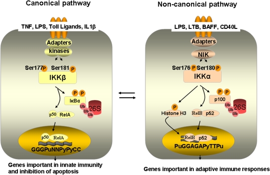
    1. Hayden MS, Ghosh S. Signaling to NF-kappaB. Genes Dev 2004;18:2195–2224. - PubMed
    1. Pantano C, Reynaert NL, van der Vliet A, Janssen-Heininger YM. Redox-sensitive kinases of the nuclear factor-kappaB signaling pathway. Antioxid Redox Signal 2006;8:1791–1806. - PubMed

Publication types