. 2009 Sep;50(9):4205-14.
doi: 10.1167/iovs.08-3214. Epub 2009 Apr 22.

Efficiency and safety of AAV-mediated gene delivery of the human ND4 complex I subunit in the mouse visual system

Affiliations

Efficiency and safety of AAV-mediated gene delivery of the human ND4 complex I subunit in the mouse visual system

John Guy et al. Invest Ophthalmol Vis Sci. 2009 Sep.

Abstract

Purpose: To evaluate the efficiency and safety of AAV-mediated gene delivery of a normal human ND4 complex I subunit in the mouse visual system.

Methods: A nuclear encoded human ND4 subunit fused to the ATPc mitochondrial targeting sequence and FLAG epitope were packaged in AAV2 capsids that were injected into the right eyes of mice. AAV-GFP was injected into the left eyes. One month later, pattern electroretinography (PERG), rate of ATP synthesis, gene expression, and incorporation of the human ND4 subunit into the murine complex I were evaluated. Quantitative analysis of ND4FLAG-injected eyes was assessed compared with green fluorescent protein (GFP)-injected eyes.

Results: Rates of ATP synthesis and PERG amplitudes were similar in ND4FLAG- and GFP-inoculated eyes. PERG latency was shorter in eyes that received ND4FLAG. Immunoprecipitated murine complex I gave the expected 52-kDa band of processed human ND4FLAG. Confocal microscopy revealed perinuclear expression of FLAG colocalized with mitochondria-specific fluorescent dye. Transmission electron microscopy revealed FLAG immunogold within mitochondria. Compared with Thy1.2-positive retinal ganglion cells (RGCs), quantification was 38% for FLAG-positive RGCs and 65% for GFP-positive RGCs. Thy1.2 positive-RGC counts in AAV-ND4FLAG were similar to counts in control eyes injected with AAV-GFP.

Conclusions: Human ND4 was properly processed and imported into the mitochondria of RGCs and axons of mouse optic nerve after intravitreal injection. Although it had approximately two-thirds the efficiency of GFP, the expression of normal human ND4 in murine mitochondria did not induce the loss of RGCs, ATP synthesis, or PERG amplitude, suggesting that allotopic ND4 may be safe for the treatment of patients with Leber hereditary optic neuropathy.


Figures

FIGURE 1
FIGURE 1
(A) Agarose gel electrophoresis of a positive control AAV-pTR plasmid containing the nuclear encoded ND4 shows the expected 242-bp band (top, lane 1). This band was absent in the retina (top, lane 2) and optic nerve (top, lane 3) of control eyes that were injected with AAV-GFP. In contrast, the RT-PCR product obtained from the RNA extracted from the retina and optic nerves of eyes injected with AAV-P1ND4FLAG revealed the expected 242-bp band, indicating in vivo transcription of the nuclear encoded human ND4 (top, lanes 4, 5). The housekeeping β-actin PCR product was seen in all ocular tissues (bottom, lanes 2-5), but it was absent with plasmid ND4FLAG serving as the template (bottom, lane 1). (B) Western blot analysis of immunoprecipitated murine complex I reacted with anti-FLAG M2 antibody revealed specific bands at 52 kDa for both the retina and the optic nerve of eyes that received intravitreal injection of AAV containing the human nuclear encoded ND4FLAG gene (B, lanes 3, 4). Western blot analysis of immunoprecipitated murine complex I extracted from the retina and optic nerve of control eyes inoculated with AAV-GFP were negative for the ND4FLAG fusion protein (B, lanes 1, 2). These findings suggest that the nuclear encoded human ND4 complex I subunit was incorporated into the mouse complex I. p-ND4, AAV plasmid containing the human ND4 gene; GFP-RET, GFP-inoculated retina; GFP-ON, GFP-inoculated optic nerve; ND4-RET, ND4-inoculated retina; ND4-ON, ND4-inoculated optic nerve.
FIGURE 2
FIGURE 2
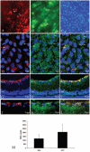
(A) Retinal flatmount from an eye injected with AAV-ND4FLAG revealed a perinuclear pattern of ND4FLAG immunofluorescence (arrows). (B) Retinal flatmount of a control eye injected with AAV-GFP showed a cytoplasmic and nuclear pattern of GFP immunofluorescence in RGCs (arrows) and along their axonal bundles of the nerve fiber layer. (C) Flatmount counterstained with DAPI shows the RGC nuclei. (D) Confocal microscopy showed the punctate and perinuclear pattern of ND4FLAG immunofluorescence (arrows) surrounding RGC nuclei counterstained with DAPI appeared similar to the epifluorescence of a bona fide mitochondrial marker (mitochondria-specific fluorescent dye; arrows, E). (F) Merged panel shows RGCs with colocalization of ND4FLAG and the mitochondrial marker (arrows). (G) Low-power longitudinal cryostat sections revealed ND4FLAG immunofluorescence was confined to the RGC layer. Mitochondria-specific fluorescent dye accumulated in RGCs (H) and colocalized with ND4FLAG (I). (J) Higher power magnification revealed ND4FLAG immunofluorescence in RGCs, (K) mitochondria-specific fluorescent dye accumulation in RGC mitochondria, and (L) colocalization of mitochondria-specific fluorescent dye with ND4FLAG. Retinal nuclei were counterstained by DAPI (D-L). (M) Bar plot shows quantitative analysis of ND4FLAG- and GFP-positive cells. RGC, retinal ganglion cell layer; INL, inner nuclear layer.
FIGURE 3
FIGURE 3
(A) Cross-sectional montaged images of the optic nerve head and peripapillary retina from the acquired 3D OCT data set of an AAV-ND4-inoculated mouse eye and the AAV-GFP-injected eye (B) show normal retinal anatomy. The OCT image is displayed in grayscale, with darker readings corresponding to lower backscattering (inner nuclear [IN] and outer nuclear [ON] layers) and brighter regions representing higher backscattering (RPE and choroid). RGC layers of experimental and control eyes were clearly recognizable. The optic nerve head of AAV-ND4 and control eyes showed no evidence of displacement of the peripapillary retina. With the use of algorithms for 3D segmentation, we measured the distance from the RGC layer to the inner boundary of the IN layer providing a geometric plot in three dimensions for the AAV-ND4 (C) and AAV-GFP (D) control eyes. Superior, inferior, nasal, and temporal quadrants are marked. The peak represents an artifact created by the measurement process and the vessels overlying the optic nerve head. Scale bar values are in millimeters. RPE, retinal pigment epithelium; ONH, optic nerve head.
FIGURE 4
FIGURE 4
(A) Excised retina was laid out flat with the RGC layer facing upward. (B) Phase-contrast light micrograph focused on the RGC layer. AAV-ND4FLAG-inoculated (C) and AAV-GFP-inoculated (D) eyes show numerous labeled Thy1.2-positive RGCs (arrows). Toluidine blue sections of AAV-ND4FLAG-inoculated (E) or AAV-GFP-inoculated (F) eyes showed no evidence of swelling of the optic nerve head. RGCs of AAV-ND4FLAG-injected (G) or AAV-GFP-injected (H) eyes showed the absence of chromatolysis or loss. (I) Bar plot of Thy1.2-positive cells shows no significant differences in RGC counts between AAV-ND4FLAG- and AAV-GFP-infected eyes.
FIGURE 5
FIGURE 5
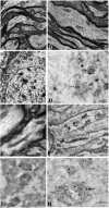
(A) Axons of the optic nerve obtained from an eye of a mouse that received no intraocular injection, and that of an AAV-ND4FLAG-inoculated eye (B) revealed normal structure of mitochondrial cristae (arrow) and axonal microtubules. (C) RGC of an AAV-ND4FLAG-injected eye exhibited a normal-appearing elliptical nucleus with pale chromatin and mitochondria with normal morphology of cristae (arrow). (D) Internalized NDFLAG-labeled immunogold (arrows) is seen within RGC mitochondria of a resin-embedded mouse retina. (E) High magnification of mitochondria (arrow) of axons of the optic nerve from an AAV-ND4FLAG-inoculated eye revealed normal cristae. (F) Optic nerve axonal mitochondria show ND4FLAG immunogold (arrows) within the organelles. (G, H) Double staining shows 6-nm anti-FLAG immunogold (small arrow) colocalizes with the larger 10-nm anti-MnSOD (large arrow) within mitochondria.
FIGURE 6
FIGURE 6
(A) Representative PERG of an ND4FLAG-inoculated eye and (B) PERG of the control eye of an animal injected with AAV-GFP. (C) Bar plot shows PERG amplitudes of ND4-inoculated eyes similar to those of GFP-inoculated eyes. (D) Bar plot of PERG latency shows a reduction in ND4-inoculated eyes compared with GFP controls.
FIGURE 7
FIGURE 7
Bar plot shows optic nerve ATP synthesis in AAV-ND4 - and AAV-GFP-inoculated eyes are comparable.

References

    1. Wallace DC, Singh G, Lott MT, et al. Mitochondrial DNA mutation associated with Leber hereditary optic neuropathy. Science. 1988;242:1427–1430. - PubMed
    1. Esposito LA, Melov S, Panov A, et al. Mitochondrial disease in mouse results in increased oxidative stress. Proc Natl Acad Sci USA. 1999;96:4820–4825. - PMC - PubMed
    1. Carelli V, Ghelli A, Bucchi L, et al. Biochemical features of mtDNA 14484 (ND6/M64V) point mutation associated with Leber’s hereditary optic neuropathy. Ann Neurol. 1999;45:320–328. - PubMed
    1. Glick B, Schatz G. Import of proteins into mitochondria. Annu Rev Genet. 1991;25:21–44. - PubMed
    1. Neupert W. Protein import into mitochondria. Annu Rev Biochem. 1997;66:863–917. - PubMed

Publication types

MeSH terms