. 2009 Jul 8;28(13):1867-77.
doi: 10.1038/emboj.2009.115. Epub 2009 Apr 23.

S5a promotes protein degradation by blocking synthesis of nondegradable forked ubiquitin chains

Affiliations

S5a promotes protein degradation by blocking synthesis of nondegradable forked ubiquitin chains

Hyoung Tae Kim et al. EMBO J. .

Abstract

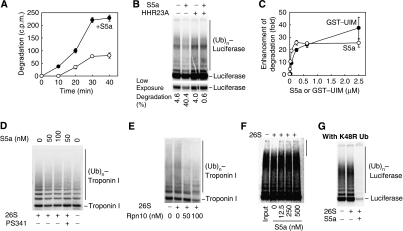
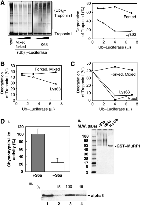
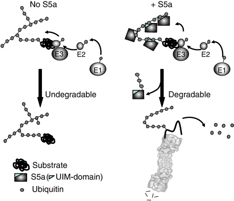
Ubiquitin (Ub)-protein conjugates formed by purified ring-finger or U-box E3s with the E2, UbcH5, resist degradation and disassembly by 26S proteasomes. These chains contain multiple types of Ub forks in which two Ub's are linked to adjacent lysines on the proximal Ub. We tested whether cells contain factors that prevent formation of nondegradable conjugates and whether the forked chains prevent proteasomal degradation. S5a is a ubiquitin interacting motif (UIM) protein present in the cytosol and in the 26S proteasome. Addition of S5a or a GST-fusion of S5a's UIM domains to a ubiquitination reaction containing 26S proteasomes, UbcH5, an E3 (MuRF1 or CHIP), and a protein substrate, dramatically stimulated its degradation, provided S5a was present during ubiquitination. Mass spectrometry showed that S5a and GST-UIM prevented the formation of Ub forks without affecting synthesis of standard isopeptide linkages. The forked Ub chains bind poorly to 26S proteasomes unlike those synthesized with S5a present or linked to Lys63 or Lys48 chains. Thus, S5a (and presumably certain other UIM proteins) function with certain E3/E2 pairs to ensure synthesis of efficiently degraded non-forked Ub conjugates.


Figures

Figure 1
Figure 1
The presence of S5a during substrate ubiquitination enhances its degradation by proteasomes. (A) S5a stimulates proteasomal degradation of luciferase. 125I–luciferase (100 nM) was denatured for 10 min at 43 °C in the presence of HSP70 (150 nM). The complex of 125I–luciferase and HSP70 was incubated with CHIP (500 nM), UbcH5 (750 nM) and purified 26S proteasomes (3.6 nM) in the presence or absence of 50 nM S5a. At different time intervals, degradation of luciferase was assayed at 37°C by measuring the appearance of TCA-soluble radioactivity with a γ-counter. (B) Addition of S5a, but not the UBL–UBA protein HHR23A stimulated Ub-dependent degradation of 125I–luciferase by 26S proteasomes. HHR23A also inhibited the enhancement of proteolysis by S5a. Purified 26S proteasomes, CHIP, UbcH5 plus S5a or HHR23A (500 nM each) were present from the start of the reaction as in (A). Ubiquitination and proteasomal degradation of 125I–luciferase were measured after 1 h using SDS–PAGE and a phosphorimager. (C) Ub-dependent degradation of 125I–luciferase increased dramatically upon addition of increasing amounts of S5a (○) or a GST fusion with the UIM domain of S5a (residues 203–329), GST–UIM (•). The reaction was carried out for 1 h and assayed as in (A), but using 125I–luciferase (250 nM), 26S proteasomes (3.6 nM), and S5a or GST–UIM were present from the outset. (D) Troponin I ubiquitinated by MuRF1 and UbcH5 in the presence of S5a (50 or 100 nM) is degraded more rapidly by 26S proteasomes. Troponin I (100 nM) was ubiquitinated by MuRF1 (500 nM) and UbcH5 (250 nM) in the presence of 26S proteasomes (3.6 nM) for 1 h at 37 °C. Degradation of troponin I was assayed by western blotting. As controls, reactions were incubated without 26S proteasomes or with 26S proteasomes preincubated with PS341 (1 μM). (E) Increasing amounts of Rpn10, the S. cerevisiae homolog of S5a, enhanced proteasomal degradation of troponin I by 26S proteasomes purified from rabbit muscle. The reaction was carried out and assayed as in (D) except that Rpn10 was used instead of human S5a. (F) Addition of S5a after the ubiquitination reaction does not enhance proteasomal degradation of ubiquitinated luciferase. Ubiquitination was carried out for 1 h at 37 °C in the absence of S5a with CHIP and UbcH5, and the Ub conjugates generated were isolated as described in the Methods. Degradation of the Ub-conjugated luciferase by 26S proteasomes (3.6 nM) was then measured at 37 °C for 1 h in the presence of increasing amounts of S5a. (G) Lys48 is not necessary for the stimulation of proteolysis by S5a. Luciferase was ubiquitinated by CHIP and UbcH5 with K48R mutant Ub in the presence of 26S proteasomes. Addition of S5a (500 nM) at the outset enhanced hydrolysis of luciferase, whereas luciferase ubiquitinated without S5a was not degraded (as was found with wild type Ub (B)).
Figure 2
Figure 2
The presence of S5a during ubiquitination enhances deubiquitination and degradation of Ub conjugates formed only by UbcH5. (A) The ubiquitination of 125I–luciferase was carried out with or without S5a (500 nM) present for 1 h at 37 °C. After this reaction was terminated, Ub conjugates were isolated by immunoprecipitation and then incubated with 3.6 nM of purified rabbit 26S proteasomes for 1 h at 37 °C. The isopeptidase caused a decrease in the amount of high-molecular-weight conjugates (higher than 191 kDa), which were synthesized with S5a present than in the conjugates synthesized without S5a. The addition of S5a (500 nM) after ubiquitination did not promote deubiquitination by the proteasome. Ubi: Ubiquitination. (B) Addition of S5a (50 or 500 nM) during ubiquitination did not enhance the proteasomal degradation of troponin I linked to homogeneous Lys48 or Lys63 Ub chains. Ubiquitination of troponin I by MuRF1 and UbcH1 from the Lys48 chain or UbcH13/Uev1a to form the Lys63 chain were carried out as in the Figure 1D. (C) At standard concentration of S5a (500 nM), only WT Ub stimulated significantly degradation of luciferase. No significant increase in degradation with S5a was seen with the K6, K11, K27, K29, K48 and K63 single-lysine mutant Ub (data not shown). However, at low concentration of S5a (25 nM), a small but reproducible increase in Ub-dependent proteolysis was seen with K33 single lysine Ub. The reactions were carried out as in Figure 1C. Insert shows enhanced hydrolysis of 125I–luciferase with low concentrations of S5a.
Figure 3
Figure 3
26S proteasomes had a lower affinity for luciferase linked to forked Ub chains than for luciferase linked to homogeneous Lys63 chains or non-forked chains synthesized in the presence of S5a. (A) Unlike Lys63 Ub–luciferase conjugates, mixed forked Ub–luciferase conjugates failed to inhibit proteasomal degradation of Lys63 Ub–troponin I conjugates, even at high concentrations. Troponin I linked to a Lys63-Ub chain (formed by MuRF1 and UbcH13/Uev1a as in Figure 2C) was isolated, and degradation by proteasomes was assayed with increasing amounts of luciferase linked to a mixed forked Ub chain (formed by CHIP and UbcH5 as in Figure 2A) or luciferase linked to a Lys63 Ub chain (formed by CHIP and UbcH13/Uev1a) in a similar manner. The degradation reaction was run for 1 h at 37 °C. Left panel shows the conjugates assayed with an anti-troponin I antibody. Right panel; percent degradation of Ub–troponin I was quantitated using a densitometer. (B) Unlike Lys63 Ub–luciferase conjugates, mixed forked Ub–luciferase conjugates failed to inhibit proteasomal degradation of Lys48 Ub–troponin I conjugates, even at high concentrations. The experiment was carried out as in (A), but troponin I was linked to a Lys48 Ub chain by MuRF1 and UbcH1 (formed as in Figure 2C). (C) The presence of S5a during ubiquitination of luciferase with UbcH5 and CHIP enhances affinity of the Ub conjugates for the proteasome, as shown by their ability to competitively inhibit degradation of Lys63 Ub–Troponin I conjugates. The capacity of different types of Ub–luciferase conjugates to inhibit proteasomal degradation of troponin I formed by MuRF1 and UbcH13/Uev1a was measured as in (A). Increasing amounts of luciferase linked to a forked Ub chain (formed by CHIP and UbcH5), a mixed Ub-chain lacking forks (formed by CHIP and UbcH5 with 500 nM of S5a) or a Lys63 Ub-chain (formed by CHIP and UbcH13/Uev1a), were added to reactions as in (A). The amount of Ub–troponin I conjugates remaining were measured using a densitometer. (D) Forked Ub chain has low affinity for 26S proteasomes. GST–MuRF1 bound to a glutathione-resin was incubated with E1 and UbcH5 to allow auto-ubiquitination with or without S5a present. After ubiquitination, other components of the reaction were removed by extensive washing. SDS–PAGE and silver-staining showed the purity of (Ub)n-GST–MURF1 after purification (panel ii). An equivalent amount of ubiquitinated GST–MuRF1 from each reaction was incubated with 26S proteasomes for 30 min at 4 °C. 26S proteasomes not bound to ubiquitinated GST–MuRF1 were removed by extensive washing. To measure bound particles, the chymotrypsin-like activity (suc–LLVY–amc cleavage) of 26S proteasomes bound to ubiquitinated GST–MuRF1 was measured using flourometer (panel i). The activity of 26S proteasomes bound to GST–MuRF1 ubiquitinated with S5a present was designated as 100%. To verify that the activity of 26S proteasomes shown in panel (i) represents the real amount of proteasomes, the same reactions were analyzed by western blotting (panel iii). The amount of α3 was measured by western blotting using an anti-α3 antibody (panel iii). Lane 1: input (1/10), Lane 2: No Ub, Lane 3: +S5a an Lane 4: without S5a. Amount of α3 from the reaction with Ub–MuRF1 ubiqutinated with S5a was set as 100%.
Figure 4
Figure 4
Proposed mechanism on how S5a prevents the formation of forked polyUb chains. Without S5a, ring-finger and U-box Ub ligases with UbcH5 form non-degradable Ub conjugates containing forked polyUb chains, because of the reaction of a lysine on a proximal Ub with a highly reactive Ub released from UbcH5. The resulting Ub conjugate is poorly degraded by proteasomes because of the presence of forks in the polyUb chain which reduces binding to the 26S proteasome. S5a binds to the growing polyUb chain and blocks the available lysines except those on the terminal Ub and then shields the chain so as to prevent the formation of more than one isopeptide linkage on one Ub moiety. During this process, the bound S5a is itself ubiquitinated.

Comment in

  • Nonconformity in ubiquitin compliance.
    Ziv I, Kleifeld O, Glickman M. Ziv I, et al. EMBO J. 2009 Jul 8;28(13):1825-7. doi: 10.1038/emboj.2009.132. EMBO J. 2009. PMID: 19587678 Free PMC article. No abstract available.

References

    1. Chau V, Tobias JW, Bachmair A, Marriott D, Ecker DJ, Gonda DK, Varshavsky A (1989) A multiubiquitin chain is confined to specific lysine in a targeted short-lived protein. Science 243: 1576–1583 - PubMed
    1. Chen L, Madura K (2002) Rad23 promotes the targeting of proteolytic substrates to the proteasome. Mol Cell Biol 22: 4902–4913 - PMC - PubMed
    1. Chuang SM, Chen L, Lambertson D, Anand M, Kinzy TG, Madura K (2005) Proteasome-mediated degradation of cotranslationally damaged proteins involves translation elongation factor 1A. Mol Cell Biol 25: 403–413 - PMC - PubMed
    1. Ciechanover A, Brundin P (2003) The ubiquitin proteasome system in neurodegenerative diseases: sometimes the chicken, sometimes the egg. Neuron 40: 427–446 - PubMed
    1. Crosas B, Hanna J, Kirkpatrick DS, Zhang DP, Tone Y, Hathaway NA, Buecker C, Leggett DS, Schmidt M, King RW, Gygi SP, Finley D (2006) Ubiquitin chains are remodeled at the proteasome by opposing ubiquitin ligase and deubiquitinating activities. Cell 127: 1401–1413 - PubMed

Publication types

MeSH terms