

Young's modulus of elasticity of Schlemm's canal endothelial cells
- PMID: 19387710
- PMCID: PMC4319365
- DOI: 10.1007/s10237-009-0156-3
Young's modulus of elasticity of Schlemm's canal endothelial cells
Abstract
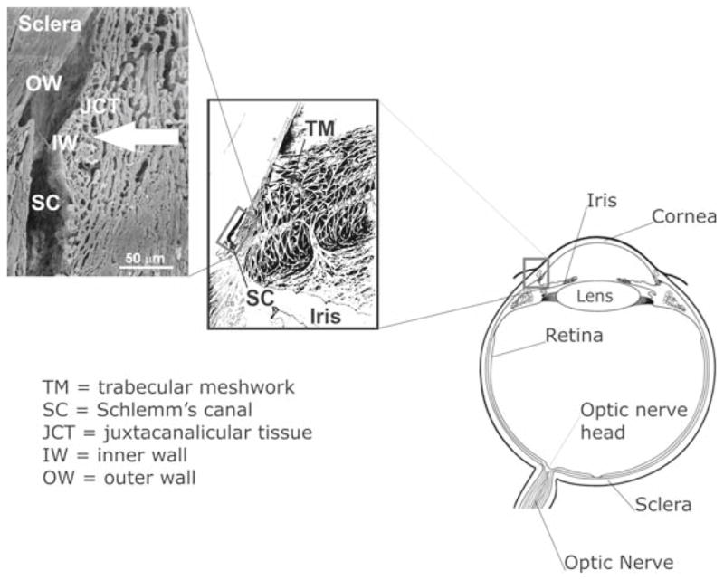
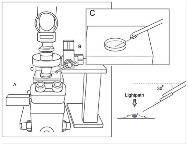
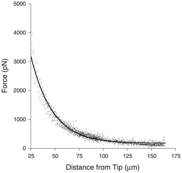
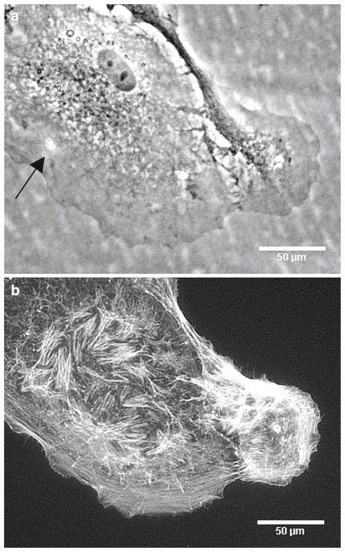
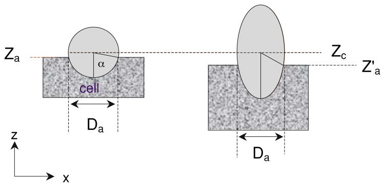
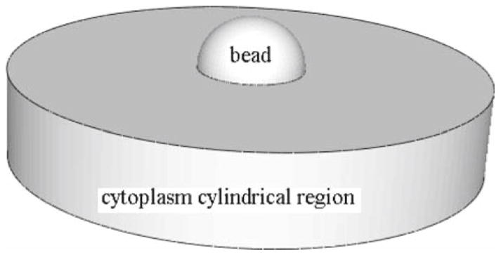
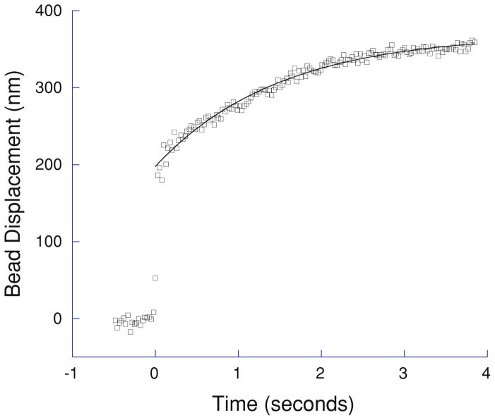
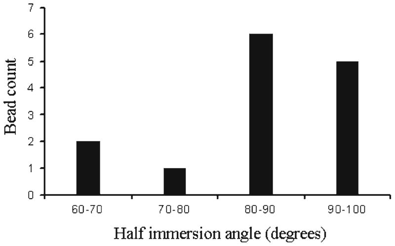
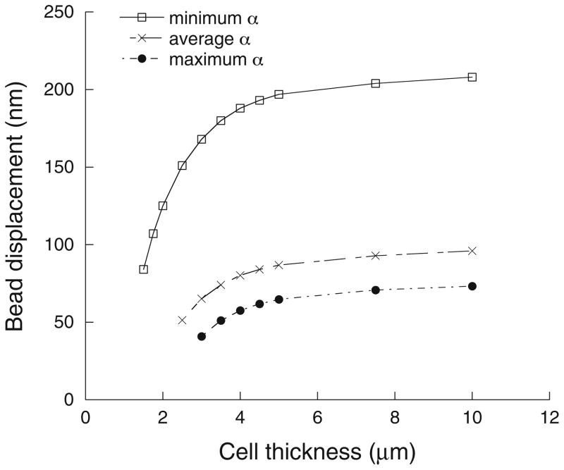
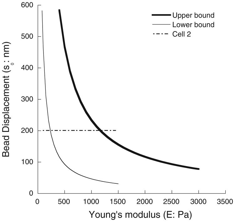
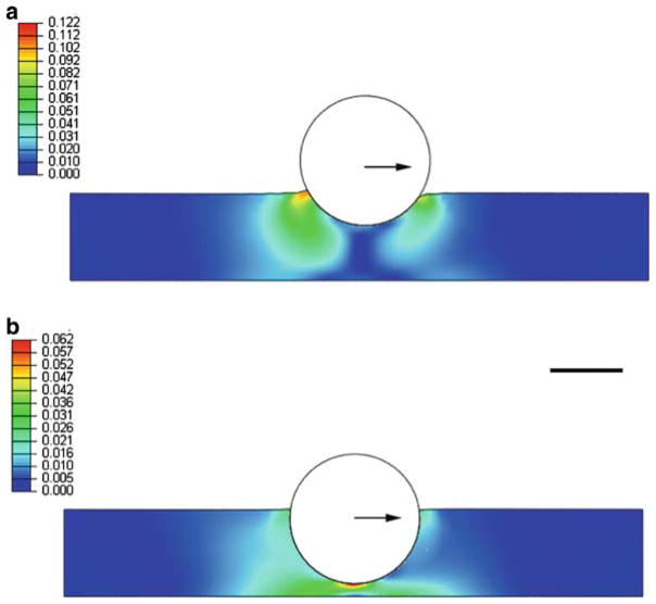
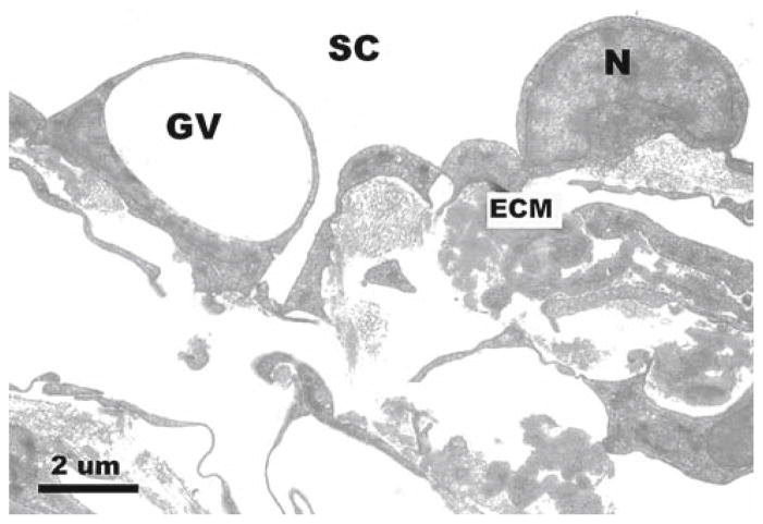
Schlemm's canal (SC) endothelial cells are likely important in the physiology and pathophysiology of the aqueous drainage system of the eye, particularly in glaucoma. The mechanical stiffness of these cells determines, in part, the extent to which they can support a pressure gradient and thus can be used to place limits on the flow resistance that this layer can generate in the eye. However, little is known about the biomechanical properties of SC endothelial cells. Our goal in this study was to estimate the effective Young's modulus of elasticity of normal SC cells. To do so, we combined magnetic pulling cytometry of isolated cultured human SC cells with finite element modeling of the mechanical response of the cell to traction forces applied by adherent beads. Preliminary work showed that the immersion angles of beads attached to the SC cells had a major influence on bead response; therefore, we also measured bead immersion angle by confocal microscopy, using an empirical technique to correct for axial distortion of the confocal images. Our results showed that the upper bound for the effective Young's modulus of elasticity of the cultured SC cells examined in this study, in central, non-nuclear regions, ranged between 1,007 and 3,053 Pa, which is similar to, although somewhat larger than values that have been measured for other endothelial cell types. We compared these values to estimates of the modulus of primate SC cells in vivo, based on images of these cells under pressure loading, and found good agreement at low intraocular pressure (8-15 mm Hg). However, increasing intraocular pressure (22-30 mm Hg) appeared to cause a significant increase in the modulus of these cells. These moduli can be used to estimate the extent to which SC cells deform in response to the pressure drop across the inner wall endothelium and thereby estimate the extent to which they can generate outflow resistance.
Figures
Similar articles
-
Finite element analysis of the pressure-induced deformation of Schlemm's canal endothelial cells.Biomech Model Mechanobiol. 2015 Aug;14(4):851-63. doi: 10.1007/s10237-014-0640-2. Epub 2014 Dec 17. Biomech Model Mechanobiol. 2015. PMID: 25516410 Free PMC article.
-
High-resolution modeling of aqueous humor dynamics in the conventional outflow pathway of a normal human donor eye.Comput Methods Programs Biomed. 2025 Mar;260:108538. doi: 10.1016/j.cmpb.2024.108538. Epub 2024 Nov 29. Comput Methods Programs Biomed. 2025. PMID: 39644783
-
Biomechanics of Schlemm's canal endothelium and intraocular pressure reduction.Prog Retin Eye Res. 2015 Jan;44:86-98. doi: 10.1016/j.preteyeres.2014.08.002. Epub 2014 Sep 16. Prog Retin Eye Res. 2015. PMID: 25223880 Free PMC article. Review.
-
Modeling the biomechanics of the conventional aqueous outflow pathway microstructure in the human eye.Comput Methods Programs Biomed. 2022 Jun;221:106922. doi: 10.1016/j.cmpb.2022.106922. Epub 2022 May 29. Comput Methods Programs Biomed. 2022. PMID: 35660940 Free PMC article.
-
The vital role for nitric oxide in intraocular pressure homeostasis.Prog Retin Eye Res. 2021 Jul;83:100922. doi: 10.1016/j.preteyeres.2020.100922. Epub 2020 Nov 28. Prog Retin Eye Res. 2021. PMID: 33253900 Free PMC article. Review.
Cited by
-
Mechanical responsiveness of the endothelial cell of Schlemm's canal: scope, variability and its potential role in controlling aqueous humour outflow.J R Soc Interface. 2012 Jun 7;9(71):1144-55. doi: 10.1098/rsif.2011.0733. Epub 2011 Dec 14. J R Soc Interface. 2012. PMID: 22171066 Free PMC article.
-
Finite element analysis of the pressure-induced deformation of Schlemm's canal endothelial cells.Biomech Model Mechanobiol. 2015 Aug;14(4):851-63. doi: 10.1007/s10237-014-0640-2. Epub 2014 Dec 17. Biomech Model Mechanobiol. 2015. PMID: 25516410 Free PMC article.
-
Aqueous humor outflow: dynamics and disease.Invest Ophthalmol Vis Sci. 2015 May;56(5):2993-3003. doi: 10.1167/iovs.15-16744. Invest Ophthalmol Vis Sci. 2015. PMID: 26024085 Free PMC article. Review. No abstract available.
-
Illuminating human health through cell mechanics.Swiss Med Wkly. 2013 Mar 14;143:w13766. doi: 10.4414/smw.2013.13766. eCollection 2013. Swiss Med Wkly. 2013. PMID: 23519500 Free PMC article. Review.
-
Synthetic Capillaries to Control Microscopic Blood Flow.Sci Rep. 2016 Feb 24;6:21885. doi: 10.1038/srep21885. Sci Rep. 2016. PMID: 26905751 Free PMC article.
References
-
- Allingham RR, de Kater AW, Ethier CR, Anderson PJ, Hertzmark E, Epstein DL. The relationship between pore density and outflow facility in human eyes. Invest Ophthalmol Vis Sci. 1992;33(5):1661–1669. - PubMed
-
- Alvarado JA, Alvarado RG, Yeh RF, Franse-Carman L, Marcellino GR, Brownstein MJ. A new insight into the cellular regulation of aqueous outflow:how trabecular meshwork endothelial cells drive a mechanism that regulates the permeability of Schlemm’s canal endothelial cells. Br J Ophthalmol. 2005;89(11):1500–1505. doi: 10.1136/bjo.2005.081307. - DOI - PMC - PubMed
-
- Bill A, Svedbergh B. Scanning electron microscopic studies of the trabecular meshwork and the canal of Schlemm—an attempt to localize the main resistance to outflow of aqueous humor in man. Acta Ophthalmol (Copenh) 1972;50:295–320. - PubMed
-
- Braet F, Rotsch C, Wisse E, Radmacher M. Comparison of fixed and living liver endothelial cells by atomic force microscopy. Appl Phys A Mater Sci Process. 1998;66:S575–S578.
Publication types
MeSH terms
Grants and funding
LinkOut - more resources
Full Text Sources
