

Comparison of piece-wise linear, linear, and nonlinear atlas-to-patient warping techniques: analysis of the labeling of subcortical nuclei for functional neurosurgical applications
- PMID: 19387981
- PMCID: PMC6871223
- DOI: 10.1002/hbm.20780
Comparison of piece-wise linear, linear, and nonlinear atlas-to-patient warping techniques: analysis of the labeling of subcortical nuclei for functional neurosurgical applications
Abstract
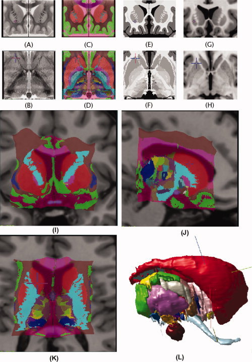
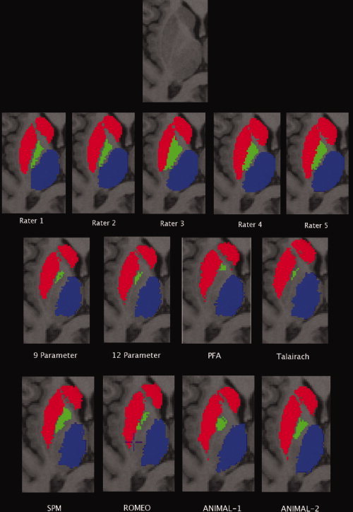
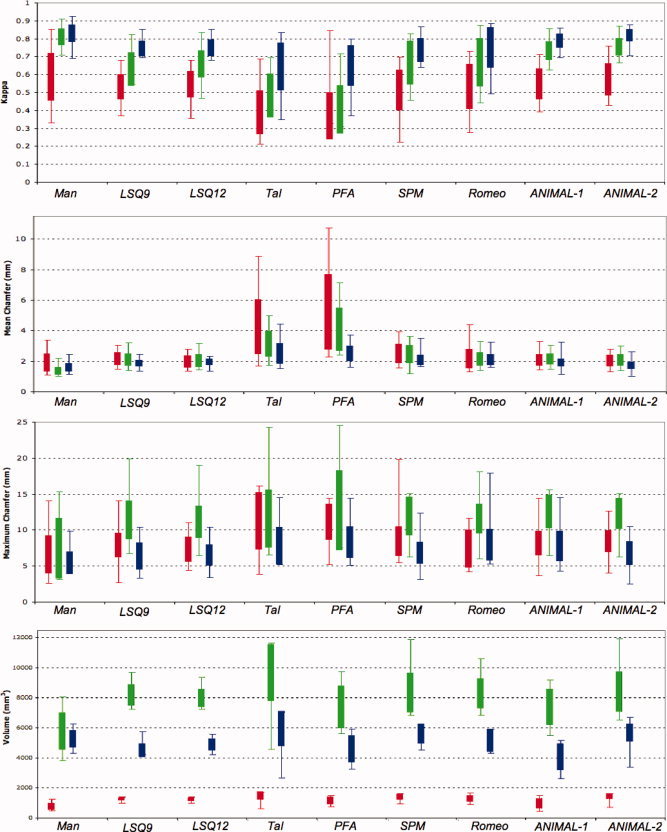
Digital atlases are commonly used in pre-operative planning in functional neurosurgical procedures performed to minimize the symptoms of Parkinson's disease. These atlases can be customized to fit an individual patient's anatomy through atlas-to-patient warping procedures. Once fitted to pre-operative magnetic resonance imaging (MRI) data, the customized atlas can be used to plan and navigate surgical procedures. Linear, piece-wise linear and nonlinear registration methods have been used to customize different digital atlases with varying accuracies. Our goal was to evaluate eight different registration methods for atlas-to-patient customization of a new digital atlas of the basal ganglia and thalamus to demonstrate the value of nonlinear registration for automated atlas-based subcortical target identification in functional neurosurgery. In this work, we evaluate the accuracy of two automated linear techniques, two piece-wise linear techniques (requiring the identification of manually placed anatomical landmarks), and four different automated nonlinear atlas-to-patient warping techniques (where two of the four nonlinear techniques are variants of the ANIMAL algorithm). Since a gold standard of the subcortical anatomy is not available, manual segmentations of the striatum, globus pallidus, and thalamus are used to derive a silver standard for evaluation. Four different metrics, including the kappa statistic, the mean distance between the surfaces, the maximum distance between surfaces, and the total structure volume are used to compare the warping techniques. The results show that nonlinear techniques perform statistically better than linear and piece-wise linear techniques. In addition, the results demonstrate statistically significant differences between the nonlinear techniques, with the ANIMAL algorithm yielding better results.
Figures
References
-
- Atkinson J,Collins DL,Bertrand G,Peters TM,Pike B,Sadikot AF 2002. Optimal location of thalamotomy lesions for tremor associated with Parkinson's disease: a probabilistic analysis based on postoperative magnetic resonance imaging and integrated digital atlas. J Neurosurg 96: 854–866. - PubMed
-
- Bardinet E,Dormont D,Malandain G,Bhattacharjee M,Pidoux B,Saleh C,Cornu P,Ayache N,Agid Y,Yelnik J ( 2005): Retrospective cross‐evaluation of a histological and deformable 3D atlas of the basal ganglia on series on Parkinsonian patients treated by deep brain stimulation In: Seventh International Conference on Medical Image Computing and Computer Assisted Intervention MICCAI 2005, Palm Springs, USA. Lecture Notes in Computer Science 3750, vol. 2 Springer; pp 385–393. - PubMed
-
- Behrens TEJ,Johansen‐Berg H,Woolrich MW,Smith SM,Wheeler‐Kingshot CAM,Boulby PA,Barker GJ,Sillery EL,Sheehan K,Ciccarelli O,Thompson AJ,Brady JM,Matthews PM ( 2003): Non‐invasive mapping of connections between human thalamus and cortex using diffusion imaging. Nat Neurosci 6: 750–757. - PubMed
-
- Bejjani BP,Dormont D,Pidoux B,Yelnik J,Damier P,Arnulf I,Bonnet AM,Marsault C,Agid Y,Philippon J,Cornu P ( 2000): Bilateral subthalamic stimulation for Parkinson's disease by using three dimensional stereotactic magnetic resonance imaging and electrophysiological guidance. J Neurosurg 92: 615–625. - PubMed
-
- Benabid AL,Koudsié A,Benazzouz A,Le Bas JF,Pollak P ( 2002): Imaging of subthalamic nucleus and ventralis intermedius of the thalamus. Movement Disorders 17: S123–S129. - PubMed
MeSH terms
LinkOut - more resources
Full Text Sources
Medical
