. 2009 May;18(5):1095-106.
doi: 10.1002/pro.119.

Key role of the N-terminus of chicken annexin A5 in vesicle aggregation

Affiliations

Key role of the N-terminus of chicken annexin A5 in vesicle aggregation

Javier Turnay et al. Protein Sci. 2009 May.

Abstract

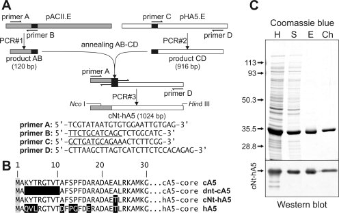
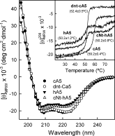
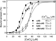
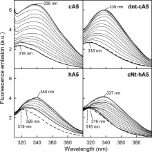
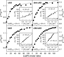
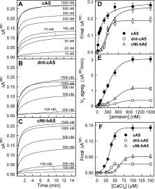
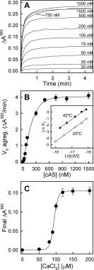
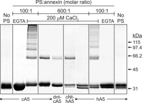
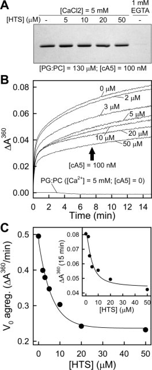
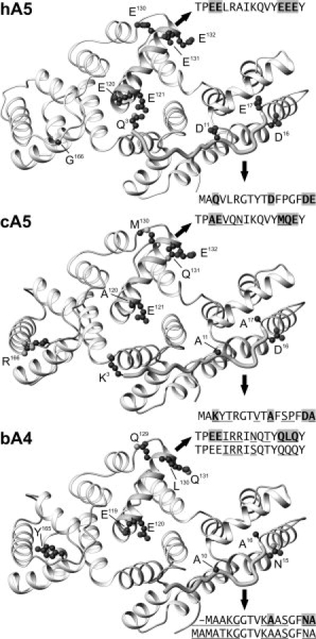
Annexins are calcium-dependent phospholipid-binding proteins involved in calcium signaling and intracellular membrane trafficking among other functions. Vesicle aggregation is a crucial event to make possible the membrane remodeling but this process is energetically unfavorable, and phospholipid membranes do not aggregate and fuse spontaneously. This issue can be circumvented by the presence of different agents such as divalent cations and/or proteins, among them some annexins. Although human annexin A5 lacks the ability to aggregate vesicles, here we demonstrate that its highly similar chicken ortholog induces aggregation of vesicles containing acidic phospholipids even at low protein and/or calcium concentration by establishment of protein dimers. Our experiments show that the ability to aggregate vesicles mainly resides in the N-terminus as truncation of the N-terminus of chicken annexin A5 significantly decreases this process and replacement of the N-terminus of human annexin A5 by that of chicken switches on aggregation; in both cases, there are no changes in the overall protein structure and only minor changes in phospholipid binding. Electrostatic repulsions between negatively charged residues in the concave face of the molecule, mainly in the N-terminus, seem to be responsible for the impairment of dimer formation in human annexin A5. Taking into account that chicken annexin A5 presents a high sequence and structural similarity with mammalian annexins absent in birds, as annexins A3 and A4, some of the physiological functions exerted by these proteins may be carried out by chicken annexin A5, even those that could require calcium-dependent membrane aggregation.


Figures

Figure 1
Figure 1
Construction and expression of the cNt-hA5 chimera. (A) Scheme of the splicing by overlap extension technique used for the construction of cNt-hA5; primers sequences are indicated. (B) Sequences of the N-terminal domains are given in the one letter code; reverse shading indicates residues that differ between cA5 and hA5. (C) Purification steps during cNt-hA5 purification followed by SDS-PAGE and Coomassie blue staining (upper panel) and Western blot using polyclonal antibodies against hA5. Exponentially growing pcNt-cA5.E transformed JA221 E. coli cultures were induced for 16 h with 1 mM IPTG, centrifuged and homogenized by sonication in the presence 2.5 mM EGTA (H); after centrifugation for 1 h at 35,000g and 4°C, the supernatant (S) was decanted and PS-enriched liposomes (1 mg/mL) and CaCl2 (2 mM final concentration) were added. Interaction with liposomes was allowed for 30 min at 4°C and the vesicles were sedimented by centrifugation, washed with buffer containing 1 mM CaCl2 and finally, the recombinant protein was extracted from the vesicles by resuspension in buffer containing 5 mM EGTA and centrifugation (E). A final ion-exchange chromatographic purification step in DEAE-cellulose yielded the cNt-hA5 preparation (Ch).
Figure 2
Figure 2
Far-UV circular dichroism spectra and thermal stability of purified recombinant proteins. Spectra were registered at 20°C in 20 mM Hepes, pH 7.4, containing 0.1M NaCl, and using protein preparations around 0.3 mg/mL. Inset: Thermal unfolding curves determined by monitorization of ellipticity changes at 208 nm between 20 and 80°C and increasing temperature at 60°C/h. Melting temperatures (Tm) are shown and correspond to mean values (±SD) of at least three independent determinations.
Figure 3
Figure 3
Liposome sedimentation assay comparing calcium dependence of the binding of recombinant proteins to PS unilamellar vesicles. Binding of recombinant proteins to 400 nm unilamellar PS vesicles was analyzed by ultracentrifugation in the presence of calcium concentrations ranging from 0 (1 mM EGTA) to 200 μM. Pellets were analyzed by SDS-PAGE followed by Coomassie blue staining and densitometry. Data represent means (±SD) of at least three independent experiments.
Figure 4
Figure 4
Fluorescence emission spectra of recombinant annexins bound to PS unilamellar vesicles. The influence of the lipid to protein molar ratio in the binding of the recombinant proteins to 50 nm PS unilamellar vesicles was analyzed by fluorescence emission spectroscopy. Spectra of the unique Trp residue present in the four recombinant proteins in the absence of calcium and PS (dashed lines) or in the presence of 200 μM CaCl2 at increasing lipid to protein molar ratio (thicker lines represent [L]/[P] = 0 and 120) are shown. The wavelengths corresponding to the maxima of the most significant spectra are shown.
Figure 5
Figure 5
Analysis of the fluorescence emission spectra of recombinant annexins bound to PS unilamellar vesicles. Spectra from Figure 4 were analyzed and the main plots show the ratio between fluorescence intensities at wavelengths corresponding to the emission maxima of completely bound or free annexin in the presence of 200 μM CaCl2. The ratio required for 50% binding ([L]/[P]50%) under these experimental conditions is indicated. Insets: plots of [L]t/[P]b vs. 1/[P]f for each recombinant protein showing the values obtained for apparent Kd and n.
Figure 6
Figure 6
Annexin-induced vesicle aggregation. (A–C) Aggregation of 100 nm unilamellar PS vesicles was studied as a function of protein concentration in the presence of 200 μM CaCl2. Aggregation was initiated by adding Ca2+ to a suspension of vesicles at the indicated protein concentration in 20 mM Hepes, pH 7.4, 0.1M NaCl, and was followed by continuously monitoring absorbance at 360 nm in a thermostatized cuvette at 20°C. hA5 did not induce aggregation even at high protein concentration (2 μM) and CaCl2 up to 1 mM. (D, E) Aggregation curves in A–C were analyzed to determine the values of final ΔA360 and apparent initial aggregation velocity (V0). The former parameter was determined from the non-linear regression to a hyperbola of the aggregation curve up to 15 min, whereas apparent V0 was determined from the analysis of the data up to only 1 min. (F) Calcium dependence of annexin-induced vesicle aggregation was carried out as previously described but maintaining constant protein concentration (100 nM for cA5 and 200 nM for dnt-cA5 and cNt-hA5) and inducing aggregation by adding Ca2+ to achieve different final concentrations (up to 200 μM). Maximum ΔA360 was determined as described above. Data in D–F correspond to mean values (±SD) of three different experiments.
Figure 7
Figure 7
cA5-induced vesicle aggregation at chicken physiological temperature. Experiments were carried out as in Figure 6 but in a thermostatized cuvette at 42°C. (A) Aggregation curves at different cA5 concentrations. (B) Analysis of the apparent initial aggregation rates; the Inset shows the double logarithmic plot of initial aggregation rates vs. cA5 concentration at lipid to protein molar ratios larger than 1000 (below 100 nM cA5). (C) Calcium dependence of cA5-induced vesicle aggregation was carried out maintaining constant protein concentration (100 nM) and inducing aggregation by adding Ca2+ to achieve different final concentrations (up to 200 μM). Data in B and C correspond to mean values (±SD) of three different experiments.
Figure 8
Figure 8
Crosslinking of annexins bound to PS unilamellar vesicles. Crosslinking of recombinant annexins was carried out at 100:1 and 600:1 phospholipid to annexin molar ratio in the presence of 200 μM Ca2+ using BS3; after stopping the crosslinking reaction, samples were centrifuged and the vesicle-bound annexin was analyzed. Controls were carried out in the presence of PS but without calcium (1 mM EGTA) or in the presence of calcium but without liposomes; in these cases, almost no annexin was sedimented and crosslinking was evaluated in aliquots from the supernatnats (“EGTA” and “No PS”).
Figure 9
Figure 9
Inhibition of cA5-induced vesicle aggregation by heparin tetrasaccharide. The effect of HTS binding in cA5-induced vesicle aggregation was assayed after preincubation of cA5 with increasing concentrations of HTS in the presence of 5 mM CaCl2 at 20°C. Afterwards, aggregation was triggered by addition of a concentrated stock of unilamellar 100 nm PG:PC liposomes. (A) Influence of HTS in the binding of cA5 to PG:PC vesicles was analyzed under identical experimental conditions to those used for the aggregation studies. 100 nm cA5-containing unilamellar vesicles were centrifuged in a Beckman Coulter Optima MAX-XP ultracentrifuge at 150,000g for 1 h at 4°C and the sediment and supernatants were analyzed by SDS-PAGE followed by Coomassie blue staining and densitometric analysis of the protein bands. A negative control without calcium (1 mM EGTA) was included. Total cA5 sedimentation was achieved in the absence of HTS and only a minor reduction in sedimentation (<7%) was observed at the highest HTS concentration used. (B) cA5-induced aggregation curves at increasing HTS concentration; no aggregation of PG:PC vesicles at 5 mM CaCl2 was observed in the absence of cA5 either in the absence or presence of HTS. (C) Analysis of the apparent initial aggregation velocity and ΔA360 after 15 min as a function of HTS concentration.
Figure 10
Figure 10
Three-dimensional structure of human and chicken annexin A5 and bovine annexin A4. X-Ray crystallography co-ordinates were obtained from the Protein Data Bank (hA5: 1ANX, residues 3–319; cA5: 1ALA, residues 3–320; bA4: 1ANN, residues 5–319). Molecules are viewed from the concave face and the lateral chains from the most significant residue changes between hA5 and cA5 are shown in a ball-and-stick style (Asp16 and Glu132 are also shown). Sequences corresponding to the N-terminal extensions and to helices IIC are shown; differences with hA5 are underlined and residues represented in a ball-and-stick style are gray-shaded. The sequences corresponding to the N-terminus and helix IIC of human annexin A4 are aligned with those from bA4. Residue numbers are assigned according to cDNA translation and thus, N-terminal methionine is Met1 even though it is not present in the wild-type proteins. The figure was prepared using the MOLMOL program.

Similar articles

Cited by

References

    1. Gerke V, Moss SE. Annexins: from structure to function. Physiol Rev. 2002;82:331–371. - PubMed
    1. Moss SE, Morgan RO. The annexins. Genome Biol. 2004;5:2.19.1–2.19.8. - PMC - PubMed
    1. Gerke V, Creutz CE, Moss SE. Annexins: linking Ca2+ signalling to membrane dynamics. Nat Rev Mol Cell Biol. 2005;6:449–461. - PubMed
    1. Turnay J, Lecona E, Guzmán-Aránguez A, Pérez-Ramos P, Fernández-Lizarbe S, Olmo N, Lizarbe MA. Annexins: structural characteristics of the N-terminus and influence and influence on the overall structure of the protein. Recent Res Dev Biochem. 2003b;4:79–95.
    1. Turnay J, Guzmán-Aránguez A, Lecona E, Pérez-Ramos P, Fernández-Lizarbe S, Olmo N, Lizarbe MA. Influence of the N-terminal domain of annexins in their functional properties. Recent Res Dev Biochem. 2003a;4:53–78.

Publication types

MeSH terms

LinkOut - more resources