. 2009 May;29(5):1190-4.
doi: 10.1002/jmri.21758.

Diminished visibility of cerebral venous vasculature in multiple sclerosis by susceptibility-weighted imaging at 3.0 Tesla

Affiliations

Diminished visibility of cerebral venous vasculature in multiple sclerosis by susceptibility-weighted imaging at 3.0 Tesla

Yulin Ge et al. J Magn Reson Imaging. 2009 May.

Abstract

Multiple sclerosis (MS) is a disease of the central nervous system characterized by widespread demyelination, axonal loss and gliosis, and neurodegeneration; susceptibility-weighted imaging (SWI), through the use of phase information to enhance local susceptibility or T2* contrast, is a relatively new and simple MRI application that can directly image cerebral veins by exploiting venous blood oxygenation. Here, we use high-field SWI at 3.0 Tesla to image 15 patients with clinically definite relapsing-remitting MS and to assess cerebral venous oxygen level changes. We demonstrate significantly reduced visibility of periventricular white matter venous vasculature in patients as compared to control subjects, supporting the concept of a widespread hypometabolic MS disease process. SWI may afford a noninvasive and relatively simple method to assess venous oxygen saturation so as to closely monitor disease severity, progression, and response to therapy.


Figures

Figure 1
Figure 1
Image post processing and segmentation of venous structures were based on SWI magnitude (A) and filtered phase (B) images. Four magnitude images (C) were manipulated with filtered phase mask by multiplication factor of 4 to create minimum intensity projection (mIP) (D). Veins, which are augmented with phase on SWI mIP image, appear as dark linear structures in contrast to brain tissue background can be segmented using statistical thresholding algorithms. Processing steps involve vascular tracking on mIP images to generate color-coded venous vasculature maps (E) with each color representing a single collecting vein and the segmentation of venous vasculature with background removal (F) to calculate the number of voxels corresponding to each vein for quantification.
Figure 2
Figure 2
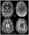
Conventional T2-weighted and SWI mIP images in a normal control (A, B) and in a patient with MS (C, D) demonstrate significantly reduced susceptibility contrast of venous blood on SWI processed mIP image in patient as opposed to healthy control subject. Note that at the periventricular level, both small (e.g. medullary veins, indicated by small arrows) and large (e.g. longitudinal caudate vein of Schlesinger, indicated by long arrow) collecting veins as well as cortical veins (arrowhead) are clearly delineated in a control participant, as opposed to their decreased visibility on SWI venograms in a patient with MS.
Figure 3
Figure 3
Segmented results of the number venous blood (number of voxels) in patients and control participants. A wider and downwards shifted distribution is found in patients with MS compared to controls and the dash (-) indicates the mean values.
Figure 4
Figure 4
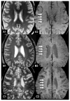
Conventional T2-weighted (A1, B1, C1) and SWI mIP images (A2, B2, C2) at the periventricular level (8mm thick) in a normal control (A1, A2) and two MS patients (B1,B2,C1,C2) demonstrate a significantly reduced number of periventricular medullary veins in patients compared to controls. MS patients with more lesions (C2) have less venous architectures depicted on SWI mIP images than patients with fewer lesions (B2).

References

    1. Filippi M. Multiple sclerosis: a white matter disease with associated gray matter damage. J Neurol Sci. 2001;185:3–4. - PubMed
    1. Brooks DJ, Leenders KL, Head G, Marshall J, Legg NJ, Jones T. Studies on regional cerebral oxygen utilisation and cognitive function in multiple sclerosis. J Neurol Neurosurg Psychiatry. 1984;47:1182–1191. - PMC - PubMed
    1. Haacke EM, Xu Y, Cheng YC, Reichenbach JR. Susceptibility weighted imaging (SWI) Magn Reson Med. 2004;52:612–618. - PubMed
    1. Poser CM, Paty DW, Scheinberg L, et al. New diagnostic criteria for multiple sclerosis: guidelines for research protocols. Ann Neurol. 1983;13:227–231. - PubMed
    1. Wilson DL, Noble JA. An adaptive segmentation algorithm for time-of-flight MRA data. IEEE Trans Med Imaging. 1999;18:938–945. - PubMed

Publication types