. 2009 Jul;11(7):1128-50.
doi: 10.1111/j.1462-5822.2009.01316.x. Epub 2009 Mar 18.

Intracellular biology and virulence determinants of Francisella tularensis revealed by transcriptional profiling inside macrophages

Affiliations

Intracellular biology and virulence determinants of Francisella tularensis revealed by transcriptional profiling inside macrophages

Tara D Wehrly et al. Cell Microbiol. 2009 Jul.

Abstract

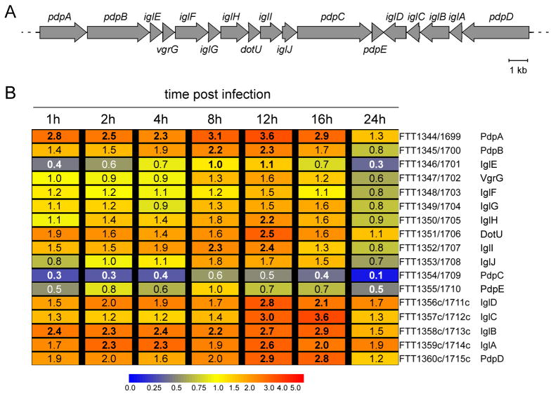
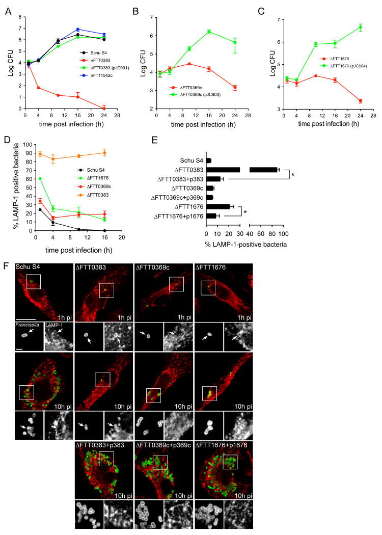
Summary The highly infectious bacterium Francisella tularensis is a facultative intracellular pathogen, whose virulence requires proliferation inside host cells, including macrophages. Here we have performed a global transcriptional profiling of the highly virulent F. tularensis ssp. tularensis Schu S4 strain during its intracellular cycle within primary murine macrophages, to characterize its intracellular biology and identify pathogenic determinants based on their intracellular expression profiles. Phagocytosed bacteria rapidly responded to their intracellular environment and subsequently altered their transcriptional profile. Differential gene expression profiles were revealed that correlated with specific intracellular locale of the bacteria. Upregulation of general and oxidative stress response genes was a hallmark of the early phagosomal and late endosomal stages, while induction of transport and metabolic genes characterized the cytosolic replication stage. Expression of the Francisella Pathogenicity Island (FPI) genes, which are required for intracellular proliferation, increased during the intracellular cycle. Similarly, 27 chromosomal loci encoding putative hypothetical, secreted, outer membrane proteins or transcriptional regulators were identified as upregulated. Among these, deletion of FTT0383, FTT0369c or FTT1676 abolished the ability of Schu S4 to survive or proliferate intracellularly and cause lethality in mice, therefore identifying novel determinants of Francisella virulence from their intracellular expression profile.


Figures

Figure 1
Figure 1
Infection cycle of F. tularensis Schu S4 within C57BL/6J murine BMMs. BMMs were infected with Schu S4 and processed at various times post infection (p.i.) for either phagosomal integrity assay or enumeration of viable intracellular bacteria (A), or for immunofluorescence or transmission electron microscopy (B-G). (A) Phagosomal escape and intracellular growth of Schu S4 within BMMs. The percentage of cytoplasmic bacteria (red curve) was determined up to 4h p.i., as described in the Materials and Methods section, and the numbers of CFUs (blue curve) were measured up to 24 h p.i. Data are means ± SD from 3 independent experiments (phagosomal integrity assay) or from a representative experiment performed in triplicate out of 3 independent repeats (CFU). (B-G) Representative confocal and electron (TEM) micrographs of intracellular Schu S4 at 30 min (B), 1 h (C), 4 h (D), 8 h (E), 12 h (F) and 24 h p.i. (G). Bacteria (appear in green) and LAMP-1-positive membranes (appear in red) were labeled. White arrowheads indicate either regions of interest in whole images or bacteria enclosed within a LAMP-1-positive compartment (panels B and G) in insets. Black arrows in TEM micrographs indicate single or double membranes surrounding intracellular bacteria (panels B and G). The asterisk in panel G indicates a bacterium enclosed within a late autophagic-like vacuole. Scale bars, 10 and 2 μm (confocal images) or 0.5 μm (TEM images)
Figure 2
Figure 2
Transcriptional profiling of intracellular Francisella (A) Representative chromatograms of total RNA isolated from either uninfected or Schu S4-infected BMMs at 0, 1, 2, 4, 8, 12, 16 and 24 h p.i., as described in the Materials and Methods section. The progressive increase in bacterial ribosomal RNAs over time mirrors the replication kinetics of intracellular bacteria (Fig. 1A). (B) Principal Component Analysis (PCA) plot showing the clustering of microarray data samples according to independent biological replicates of the same time points. PCA was performed using the Partek Genomics Suite software v6.3, as described in the Materials and Methods section. (C) Numbers of Francisella genes significantly up- or down-regulated at the various times p.i. Data from time zero samples was used as a baseline to determine genes whose mRNA levels varied by more than two fold. (D) Grouping of Francisella genes significantly up- or down-regulated according to their mRNA change index, i.e. the numbers of time points analyzed when expression of a given gene was significantly altered. Data from time zero samples was used as a baseline to determine genes whose mRNA levels varied by more than 2 fold. Numbers above brackets indicate the sums of genes in the included categories.
Figure 3
Figure 3
Differential expression profiles of Francisella genes correlate with specific intracellular stages. Color-coded representations of mRNA levels of general stress response (A), oxidative stress response (B) and iron acquisition (C) genes at all time points analyzed, showing differential expression profiles of Schu S4 genes during the macrophage infection cycle. Numbers indicate fold changes in mRNA levels relative to time zero. Bold numbers correspond to significant changes in mRNA levels, as determined by statistical analyses described in the Materials and Methods section. ND indicates undetermined values due to the lack of detectable intracellular mRNA signals. Schu S4 locus tags and encoded functions are indicated. (D) Intracellular expression profile of clpB (FTT1769c) as determined by quantitative RT-PCR. ClpB mRNAs were quantified and normalized to those of the constitutively expressed gyrA (FTT1575c) gene.
Figure 4
Figure 4
Amino acid metabolism and oligopeptide transport during the Francisella intracellular cycle. Color-coded representations of mRNA levels of amino acid metabolism (A), and di/tripeptide transporter (B) genes at all time points analyzed. Numbers indicate fold changes in mRNA levels relative to time zero. Bold numbers correspond to significant changes in mRNA levels, as determined by statistical analyses described in the Materials and Methods section. ND indicates undetermined values due to the lack of detectable intracellular mRNA signals. Schu S4 locus tags, encoded functions and corresponding protein names or homologs are indicated.
Figure 5
Figure 5
Intracellular expression profiles of FPI genes by DNA Genechip analysis. (A) Schematic representation of the FPI locus in Schu S4. The gene nomenclature is according to Ludu et al. (Ludu et al., 2008). (B) Color-coded representations of FPI gene mRNA levels during the infection cycle, showing similar expression profiles within the FPI. Numbers indicate fold changes in mRNA levels relative to time zero. Bold numbers correspond to significant changes in mRNA levels, as determined by statistical analyses described in the Materials and Methods section. Schu S4 locus tags and encoded proteins are indicated. Single probe sets for both FPI-1 and FPI-2 loci were spotted on the Genechip and identically matched with each FPI locus sequences. FPI genes are coordinately expressed and induced intracellularly.
Figure 6
Figure 6
Intracellular expression of FPI genes and proteins during the infection cycle. (A) Intracellular expression profiles of pdpD, iglA, iglC, pdpA and pdpC as determined by quantitative RT-PCR. Specific mRNAs from each gene were quantified and normalized to those of the constitutively expressed gyrA (FTT1575c) gene. Note the differences in the scale of normalized mRNA levels between genes. (B) Intrabacterial levels of IglC and PdpC during the intracellular cycle. BMMs were infected with Schu S4 as described in the Materials and Methods section and samples were processed for Western Blot analysis using either a monoclonal anti-IglC antibodies (upper panel) or polyclonal anti-PdpC antibodies (lower panels). Sample loading was normalized to intracellular CFU counts. Western blots are from a representative experiment out of 3 independent repeats.
Figure 7
Figure 7
Intracellular expression profiles of upregulated genes encoding hypothetical functions. (A) Color-coded representations of Francisella gene mRNA levels during the infection cycle, showing various profiles of intracellular upregulation. Numbers indicate fold changes in mRNA levels relative to time zero. Bold numbers correspond to significant changes in mRNA levels, as determined by statistical analyses described in the Materials and Methods section. Schu S4 locus tags and putative encoded proteins are indicated. Locus tags in blue indicate genes known to be regulated by MglA (Brotcke et al., 2006). (B) Intracellular expression profiles of FTT0989, FTT1392 and FTT1542c, as determined by quantitative RT-PCR. Specific mRNAs from each gene were quantified and normalized to those of the constitutively expressed gyrA (FTT1575c) gene.
Figure 8
Figure 8
SacB-assisted allelic replacement vector pJC84 and in-frame deletion of FTT0383, FTT0369c and FTT1676 in Schu S4. (A) Map of pJC84 (3775 bp; GenBank accession number FJ155667) showing the bicistronic aph-sacB operon under the control of the Francisella groEL promoter region. Unique restriction sites are indicated in black. (B) Schematic representation of the FTT0383 region of the Schu S4 chromosome before and after allelic replacement using pJC84ΔFTT0383 (see Supplementary Material). Hatched lines indicate the chromosomal regions flanking the FTT0383 locus that were used for allelic replacement. The allelic replacement was designed to preserve the integrity of the converging psd gene. (C) Schematic representation of the FTT0369c region of the Schu S4 chromosome before and after allelic replacement using pJC84ΔFTT0369c (see Supplementary Material). Hatched lines indicate the chromosomal regions flanking the FTT0369c locus that were used for allelic replacement. The allelic replacement was designed to preserve the integrity of the mviN gene located immediately downstream of FTT0369c. (D) Schematic representation of the FTT1676 region of the Schu S4 chromosome before and after allelic replacement using pJC84ΔFTT1676 (see Supplementary Material). Hatched lines indicate the chromosomal regions flanking the FTT1676 locus that were used for allelic replacement.
Figure 9
Figure 9
Deletion of FTT0383, FTT0369c or FTT1676 affects intracellular survival or growth of Schu S4. (A) Intracellular growth of Schu S4, its isogenic ΔFTT1542c and ΔFTT0383 mutants and the complemented ΔFTT0383[pJC901] (ΔFTT0383+p383) strain in BMMs. BMMs were infected with either strains as described in the Materials and Methods section and CFUs were enumerated at various times p.i. Data are means ± SD from a representative experiment performed in triplicate out of 2 to 4 independent repeats. (B) Intracellular growth of the ΔFTT0369c and the complemented ΔFTT0369c[pJC903] (ΔFTT0369c+p369c) strains in BMMs. BMMs were infected with either strains as described and CFUs were enumerated at various times p.i. Data are means + SD from a representative experiment performed in triplicate out of 2 independent repeats. (C) Intracellular growth of the ΔFTT1676 and the complemented ΔFTT1676[pJC904] (ΔFTT1676+p1676) strains in BMMs. BMMs were infected with either strains as described and CFUs were enumerated at various times p.i. Data are means + SD from a representative experiment performed in triplicate out of 2 independent repeats. (D) Quantitation of bacterial enclosure within phagosomal membranes. BMMs were infected for 1, 4, 10 or 16 h with either Schu S4 or its isogenic ΔFTT0383, ΔFTT0369c, and ΔFTT1676 mutants. Samples were processed for immunofluorescence labeling of bacteria and LAMP-1-positive membranes and numbers of bacteria enclosed within LAMP-1-positive compartments were scored. At least 100 bacteria per experiment were scored for each condition. Data are means ± SD from three independent experiments. (E) Quantitation of bacterial enclosure within phagosomal membranes at 4 h p.i. BMMs were infected for 4 h with either Schu S4 or its isogenic ΔFTT0383, ΔFTT0369c, and ΔFTT1676 mutants or the corresponding complemented strains. Samples were processed for immunofluorescence labeling of bacteria and LAMP-1-positive membranes and numbers of bacteria enclosed within LAMP-1-positive compartments were scored. At least 100 bacteria per experiment were scored for each condition. Data are means + SD from three independent experiments. Asterisks indicate a statistically significant difference (P<0.05, 2-tailed unpaired Student's t-test). (F) Representative confocal micrographs of BMMs infected for either 1 or 10 h p.i. with either Schu S4 or its isogenic ΔFTT0383, ΔFTT0369c, and ΔFTT1676 mutants (upper and middel panels), or for 10 h with the respective complemented strains ΔFTT0383+p383, ΔFTT0369c+p369c and ΔFTT1676+p1676 (lower panels). Samples were processed for immunofluorescence labeling of bacteria (appear in green) and LAMP-1-positive membranes (appear in red). Magnified insets show single channel images of the boxed region on the whole image. White arrowheads indicate bacteria of interest. Scale bars, 10 or 2 μm.
Figure 10
Figure 10
The FTT0383, FTT0369c and FTT1676 loci are required for Schu S4 virulence in mice. Survival curves of BALB/cJ mice infected with either Schu S4 or its isogenic ΔFTT0383, ΔFTT0369c or ΔFTT1676 mutants after intradermal (A) or intranasal (B) inoculations. Intradermal inoculums were 30 (Schu S4), 46 (ΔFTT0383), 47 (ΔFTT0369c) and 40 (ΔFTT1676) CFUs; intranasal inoculums were 8 (Schu S4), 9 (ΔFTT0383), 13 (ΔFTT0369c) and 14 (ΔFTT1676) CFUs.

Similar articles

Cited by

References

    1. Bader MW, Navarre WW, Shiau W, Nikaido H, Frye JG, McClelland M, et al. Regulation of Salmonella typhimurium virulence gene expression by cationic antimicrobial peptides. Molecular microbiology. 2003;50:219–230. - PubMed
    1. Baron GS, Nano FE. MglA and MglB are required for the intramacrophage growth of Francisella novicida. Molecular microbiology. 1998;29:247–259. - PubMed
    1. Belland RJ, Zhong G, Crane DD, Hogan D, Sturdevant D, Sharma J, et al. Genomic transcriptional profiling of the developmental cycle of Chlamydia trachomatis. Proc Natl Acad Sci U S A. 2003;100:8478–8483. - PMC - PubMed
    1. Bergman NH, Anderson EC, Swenson EE, Janes BK, Fisher N, Niemeyer MM, et al. Transcriptional profiling of Bacillus anthracis during infection of host macrophages. Infect Immun. 2007;75:3434–3444. - PMC - PubMed
    1. Boschiroli ML, Ouahrani-Bettache S, Foulongne V, Michaux-Charachon S, Bourg G, Allardet-Servent A, et al. The Brucella suis virB operon is induced intracellularly in macrophages. Proc Natl Acad Sci U S A. 2002;99:1544–1549. - PMC - PubMed

Publication types

MeSH terms

Substances

Associated data