. 2009 Apr 23:9:39.
doi: 10.1186/1472-6750-9-39.

A modified and automated version of the 'Fluorimetric Detection of Alkaline DNA Unwinding' method to quantify formation and repair of DNA strand breaks

Affiliations

A modified and automated version of the 'Fluorimetric Detection of Alkaline DNA Unwinding' method to quantify formation and repair of DNA strand breaks

María Moreno-Villanueva et al. BMC Biotechnol. .

Abstract

Background: Formation and repair of DNA single-strand breaks are important parameters in the assessment of DNA damage and repair occurring in live cells. The 'Fluorimetric Detection of Alkaline DNA Unwinding (FADU)' method [Birnboim HC, Jevcak JJ. Cancer Res (1981) 41:1889-1892] is a sensitive procedure to quantify DNA strand breaks, yet it is very tedious to perform.

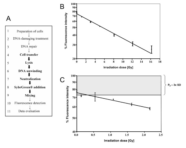
Results: In order (i) to render the FADU assay more convenient and robust, (ii) to increase throughput, and (iii) to reduce the number of cells needed, we have established a modified assay version that is largely automated and is based on the use of a liquid handling device. The assay is operated in a 96-well format, thus greatly increasing throughput. The number of cells required has been reduced to less than 10,000 per data point. The threshold for detection of X-ray-induced DNA strand breaks is 0.13 Gy. The total assay time required for a typical experiment to assess DNA strand break repair is 4-5 hours.

Conclusion: We have established a robust and convenient method measuring of formation and repair of DNA single-strand breaks in live cells. While the sensitivity of our method is comparable to current assays, throughput is massively increased while operator time is decreased.


Figures

Figure 1
Figure 1
(A) Overview of the essential steps of the modified and automated FADU assay. After preparation of the cells, DNA damage is inflicted, followed by further incubation to allow DNA repair. The automated steps are highlighted in bold. (B), (C) Dose-dependent induction of DNA strand breaks by X-rays in human PBMC (B, high dose range; C, low dose range). PBMC were exposed to various doses between 0 and 16.2 Gy. Note that the percentage of unwound DNA increased with increasing doses. Data are presented as means ± SDs from 3 independent experiments performed on PBMC from different donors. The detection threshold was set as mean minus 3 SD of the unirradiated controls (P0).
Figure 2
Figure 2
(A) Comparison of SybrGreen (Sybr) with ethidium bromide (EtBr) as a fluorescent probe for the FADU assay. For each experimental point in this experiment, 80,000 human PBMC were suspended in 70 μl suspension buffer at 0°C. For B samples, 70 μl of lysis buffer was added and the mixture was sheared by 20 passages through a 0.5-mm cannula, followed by transfer to a 96-well plate [140 μl per well]. For P and T samples 70 μl of cell suspension was transferred per well and 70 μl lysis solution was added at 0°C. This and all subsequent pipetting steps were performed by the LHD. After 12 min of lysis, 70 μl of unwinding solution was added on the top of the cell lysate followed by incubation at 15°C for 90 min. To stop DNA unwinding, neutralisation solution was added. Then 150 μl of the mixture was combined in plastic cuvettes with 500 μl of either Sybr or EtBr solution. Fluorescence detection was done at excitation 480 nm and emission 520 nm. (B) Alkaline unwinding of plasmid DNA as a model substrate. The circular form of the plasmid (white) could not be unwound and retained nearly 100% of the total fluorescence (T circular). The P0 values of the linear form (grey) represent unwound DNA and display less than 20% double/stranded DNA compared to non-denaturing control conditions (T linear). To obtain the T values, neutralisation solution was added before the alkaline solution. Error bars represent SDs from 8 replicates.
Figure 3
Figure 3
Measurement of fluorescence signals at various time points after SybrGreen addition. PBMC were exposed to an X-ray irradiation dose of 0.47 Gy (P1) or 2.3 Gy (P2). Cells in T and P0 samples were not irradiated. Note that the fluorescence signal decreased with increasing irradiation dose, as expected. Fluorescence intensity during the first 20 min upon SybrGreen addition remained constant but decreased thereafter. The ratios P0/T, P1/P0 and P2/P1 remained constant over the whole observation period.
Figure 4
Figure 4
DNA damage and repair in several different cell systems. HeLa S3 cells (A), Jurkat cells (B) and human PBMC (C) were damaged with 2.3 Gy of X-irradiation (P1). To allow repair, cells were incubated at 37°C for 40 min (R). T and P0 samples were controls. In all cases the fluorescence signal of the P1 samples was lower that the control value P0, and those of the R values higher than the damaged samples P1. Error bars represent SDs from 8 replicates.
Figure 5
Figure 5
Time course of DNA strand break repair of Jurkat cells in intervals of 5 min. Cells were X-irradiated on ice with 6.8 Gy (basal line). To measure DNA strand break repair, the damaged cells were incubated at 37°C for different time periods as indicated (grey columns). The black column represents level of SybrGreen fluorescence obtained in undamaged cells (P0 values). Error bars represent SDs from 3 replicates.

References

    1. Birnboim HC, Jevcak JJ. Fluorometric method for rapid detection of DNA strand breaks in human white blood cells produced by low doses of radiation. Cancer Res. 1981;41:1889–1892. - PubMed
    1. Kanter PM, Schwartz HS. A fluorescence enhancement assay for cellular DNA damage. Mol Pharmacol. 1982;22:145–151. - PubMed
    1. Batel R, Jaksic Z, Bihari N, Hamer B, Fafandel M, Chauvin C, Schröder HC, Müller WE, Zahn RK. A microplate assay for DNA damage determination (fast micromethod) Anal Biochem. 1999;270:195–200. doi: 10.1006/abio.1999.4098. - DOI - PubMed
    1. Elmendorff-Dreikorn K, Chauvin C, Slor H, Kutzner J, Batel R, Müller WE, Schröder HC. Assessment of DNA damage and repair in human peripheral blood mononuclear cells using a novel DNA unwinding technique. Cell Mol Biol. 1999;45:211–218. - PubMed
    1. Baumstark-Khan C, Hentschel U, Nikandrova Y, Krug J, Horneck G. Fluorometric analysis of DNA unwinding (FADU) as a method for detecting repair-induced DNA strand breaks in UV-irradiated mammalian cells. Photochem Photobiol. 2000;72:477–484. doi: 10.1562/0031-8655(2000)072<0477:FAODUF>2.0.CO;2. - DOI - PubMed

Publication types

LinkOut - more resources