. 2009;14(2):82-92.
doi: 10.1179/135100009X392520.

Hydroxyl radical oxidation of guanosine 5'-triphosphate (GTP): requirement for a GTP-Cu(II) complex

Affiliations

Hydroxyl radical oxidation of guanosine 5'-triphosphate (GTP): requirement for a GTP-Cu(II) complex

Giselle Cerchiaro et al. Redox Rep. 2009.

Abstract

Levels of oxidized guanosine base in DNA have become a hallmark biomarker in assessing oxidative stress implicated in a variety of disease and toxin-induced states. However, there is evidence that the guanosine in the nucleotide triphosphate pool (GTP) is more susceptible to oxidation than guanosine residues incorporated into nucleic acids and this causes a substantial amount of the oxidized product, 8-oxoguanosine 5'-triphosphate (oxo(8)GTP), to accumulate in cell-free and in cell-culture preparations. Electron paramagnetic resonance (EPR) spectroscopy and direct EPR analysis of free radical production by copper sulfate and L-ascorbic acid demonstrates that the hydroxyl radical (HO(*)) is produced via oxidation of Cu(+) to Cu(2+) while in a complex with GTP. This HO(*) production is dependent on the availability of oxygen and the presence of GTP in the reaction milieu. Verification of free radical-mediated production of oxo(8)GTP is presented using HPLC with electrochemical detection and matrix-assisted laser desorption/ionization linear time-of-flight mass spectrometry (MALDI-LTOF-MS). The sum of these results is presented in a novel mechanism of GTP oxidation by Cu(2+) and L-ascorbic acid. A better understanding of the chemistry involved in this oxidative modification of GTP facilitates a more comprehensive understanding of its potential physiological consequences.


Figures

Figure 1
Figure 1. EPR spectra of GTP, L-ascorbic acid, and Cu(II)
A) and B) EPR spectra of control experiments in PBS, pH =7.4, 1 mM L-ascorbic acid, and 80mM DMPO incubated for 2 min (A) and 30 min (B). C) to H) EPR spectra of reactions of 1 mM GTP, 1 mM L-ascorbic acid, 10 μM Cu(II) sulfate, and 80mM DMPO in air-saturated PBS, pH 7.4 solution. C′) to H′) EPR spectra of reactions exactly as in C to H, with the exception that the PBS pH 7.4 solution is devoid of oxygen by saturation with argon for 30 minutes. All spectra were collected at room temperature (22 °C) and plotted on the same scale (magnetic field and intensity of EPR) for comparison. EPR conditions: receiver gain 5.02 × 10⁴, modulation amplitude 1 G, time constant 163.84 ms, frequency 9.687 GHz, power 20 mW and 1024 points resolution.
Figure 2
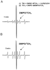
Figure 2. EPR spectra of GTP, L-ascorbic acid, and Cu(II) with DMSO
Experiment conducted in PBS, pH =7.4, 1mM L-ascorbic acid, and 80 mM DMPO incubated for 1 min (A) or 3 min (B). EPR conditions: receiver gain 5.02 × 10⁴, modulation amplitude 1 G, time constant 163.84 ms, frequency 9.687 GHz, power 20 mW and 1024 points resolution.
Figure 3
Figure 3. EPR spectra of GTP and Cu(II) or L-ascorbic acid
A) to G) EPR spectra of the reaction of 1mM GTP, 10 μM Cu(II) sulfate, and 80 mM DMPO in air-saturated PBS, pH=7.4 solution. H) to L) EPR spectra of the reaction of 1 mM GTP, 1 mM L-ascorbic acid, and 80 mM DMPO in air-saturated PBS, pH 7.4 solution. M) to P) EPR spectra of the reaction of 1mM L-ascorbic acid, 10 μM Cu(II) sulfate, and 80 mM DMPO in air-saturated PBS, pH=7.4 solution. All spectra were collected at room temperature (22 °C) and plotted on the same scale (magnetic field and intensity of EPR) for comparison. EPR conditions: receiver gain 5.02 × 10⁴, modulation amplitude 1 G, time constant 163.84 ms, frequency 9.687 GHz, power 20 mW and 1024 points resolution.
Figure 4
Figure 4. Effects of pH and reactant concentrations on free radical generation
A) and N) EPR spectra of 1 mM L-ascorbic and 80 mM DMPO in PBS, pH 7.0 (A) and PBS, pH 5.7 (N); B) to E) EPR spectra of the reaction of 1 mM GTP, 1 mM L-ascorbic acid, 10 μM Cu(II) sulfate, and 80 mM DMPO, in air-saturated PBS, pH 7.0 solution. F) to I) EPR spectra of the reaction of 2 mM GTP, 1 mM L-ascorbic acid, 30 μM Cu(II) sulfate, and 80 mM DMPO, in air-saturated PBS, pH=7.0 solution. J) to M) EPR spectra of the reaction of 3 mM GTP, 1mM L-ascorbic acid, 40 μM Cu(II) sulfate, and 80 mM DMPO, in air-saturated PBS, pH 7.0 solution. O) to P) EPR spectra of the reaction of 1 mM GTP, 1 mM L-ascorbic acid, 10 μM Cu(II) sulfate, and 80 mM DMPO, in air-saturated PBS, pH 5.7 solution. All spectra were collected at room temperature (22 °C) and plotted on the same scale (magnetic field and intensity of EPR) for comparison. EPR conditions: receiver gain 5.02 × 10⁴, modulation amplitude 1 G, time constant 163.84 ms, frequency 9.687 GHz, power 20 mW and 1024 points resolution.
Figure 5
Figure 5. Direct EPR analysis of Cu(II) and GTP
A) EPR spectrum of 2 mM Cu(II) sulfate. B) EPR spectrum of 500 μM Cu(II) sulfate with 2 mM of GTP. All solutions diluted in water, PBS, and 5% glycerol. EPR conducted at 77K.
Figure 6
Figure 6. Oxygen consumption by GTP, Cu(II), and L-ascorbic acid
Oxygen measurements using a Clark electrode of the reaction of 1 mM GTP, 1 mM L-ascorbic acid, 10 μM Cu(II) sulfate (▪) or Cu(Imine) (formula image), in air-saturated PBS, pH 7.4 solution in a total cell volume of 1.5 mL at (22.0 ± 0.2) °C.
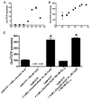
Figure 7
Figure 7. Oxo8GTP formation analysis by HPLC-EC and MALDI-TOF
A) and D) are HPLC-EC chromatogram and MALDI-TOF spectra of oxo8G (reported as oxo8GTP, see Methods) and GTP respectively, in a four-hour incubation of 1 mM GTP and 1 mM L-ascorbic acid at 37°C. Inset of D) is from a standard solution containing 1:1 GTP:Oxo8GTP (m/z GTP:522.21; m/z oxo8GTP: 539.41). B) and E) is a four-hour incubation of 1mM GTP and 200 μM Cu(II) sulfate at 37°C. C) and F) is a four-hour incubation of 1mM GTP, 1mM L-ascorbic acid, and 10 μM Cu(II) sulfate at 37°C. HPLC-EC chromatograms show traces collected at 250 mV EC channel. MALDI-TOF spectra are normalized to % Intensity of signal and baselines were corrected for noise.
Figure 8
Figure 8. Oxo8GTP in GTP, Cu(II), L-ascorbic acid and Aβ1-42 reactions
A) Oxo8GTP levels quantified by HPLC-EC in one-hour incubations of 1mM GTP and increasing concentrations of L-ascorbic acid. B) Oxo8GTP levels quantified by HPLC-EC in four-hour incubations of 1 mM GTP, 1 mM L-ascorbic acid, and increasing concentrations of Cu(II) sulfate. C) Oxo8GTP levels quantified by HPLC-EC in four-hour incubations of 1 mM GTP and 1 mM L-ascorbic acid, 200 μM Cu(II) sulfate, 1mM L-ascorbic acid/ 10 μM Cu(II) sulfate, 100 nM Aβ1-42, and 10 μM Cu(II) sulfate/100 nM Aβ1-42 at 37°C. Data represent mean ± SEM. n= 2-9. * p< 0.05 applying a one-way ANOVA with Newman-Keuls Multiple Comparison Test post-hoc analysis.
Figure 9
Figure 9. Mechanism proposed for the oxidation of GTP by Cu(II) and L-ascorbic acid

References

    1. Sies H. Role of reactive oxygen species in biological processes. Klin Wochenschr. 1991;69(2123):965–8. - PMC - PubMed
    1. Halliwell B, Gutteridge JM. Oxygen free radicals and iron in relation to biology and medicine: some problems and concepts. Arch Biochem Biophys. 1986;246(2):501–14. - PubMed
    1. Einolf HJ, Schnetz-Boutaud N, Guengerich FP. Steady-state and pre-steady-state kinetic analysis of 8-oxo-7,8-dihydroguanosine triphosphate incorporation and extension by replicative and repair DNA polymerases. Biochemistry. 1998;37(38):13300–12. - PubMed
    1. Hayakawa H, Taketomi A, Sakumi K, Kuwano M, Sekiguchi M. Generation and elimination of 8-oxo-7,8-dihydro-2′-deoxyguanosine 5′-triphosphate, a mutagenic substrate for DNA synthesis, in human cells. Biochemistry. 1995;34(1):89–95. - PubMed
    1. Russo MT, Blasi MF, Chiera F, Fortini P, Degan P, Macpherson P, et al. The oxidized deoxynucleoside triphosphate pool is a significant contributor to genetic instability in mismatch repair-deficient cells. Mol Cell Biol. 2004;24(1):465–74. - PMC - PubMed

Publication types

MeSH terms

LinkOut - more resources