. 2009 Apr 15;328(2):328-41.
doi: 10.1016/j.ydbio.2009.01.037. Epub 2009 Feb 4.

Residual microRNA expression dictates the extent of inner ear development in conditional Dicer knockout mice

Affiliations

Residual microRNA expression dictates the extent of inner ear development in conditional Dicer knockout mice

Garrett A Soukup et al. Dev Biol. .

Abstract

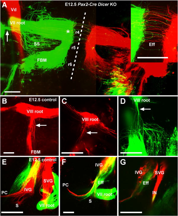
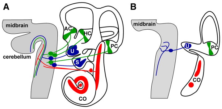
Inner ear development requires coordinated transformation of a uniform sheet of cells into a labyrinth with multiple cell types. While numerous regulatory proteins have been shown to play critical roles in this process, the regulatory functions of microRNAs (miRNAs) have not been explored. To demonstrate the importance of miRNAs in inner ear development, we generated conditional Dicer knockout mice by the expression of Cre recombinase in the otic placode at E8.5. Otocyst-derived ganglia exhibit rapid neuron-specific miR-124 depletion by E11.5, degeneration by E12.5, and profound defects in subsequent sensory epithelial innervations by E17.5. However, the small and malformed inner ear at E17.5 exhibits residual and graded hair cell-specific miR-183 expression in the three remaining sensory epithelia (posterior crista, utricle, and cochlea) that closely corresponds to the degree of hair cell and sensory epithelium differentiation, and Fgf10 expression required for morphohistogenesis. The highest miR-183 expression is observed in near-normal hair cells of the posterior crista, whereas the reduced utricular macula demonstrates weak miR-183 expression and develops presumptive hair cells with numerous disorganized microvilli instead of ordered stereocilia. The correlation of differential and delayed depletion of mature miRNAs with the derailment of inner ear development demonstrates that miRNAs are crucial for inner ear neurosensory development and neurosensory-dependent morphogenesis.


Figures

Figure 1
Figure 1
Developmental defects and miRNA depletion in Pax2-Cre Dicer KO mice. A, Overview of developmental defects. Depicted are external features of the head (left) and approximate midsagittal section (right). Notable defects exemplified by comparison of E18.5 Pax2-Cre Dicer KO and control littermates include abbreviated craniofacial development and abrogated eyelid and cerebellum (CB) development (denoted by the asterisk). B, Floxed allele recombination in Pax2-Cre control (Dicerflox/wt) and Dicer KO (Dicerflox/flox) mouse brain. Depicted is X-Gal staining indicating Pax2-Cre positive tissue in which the Rosa26-LacZ reporter (R26R) allele has been recombined to express functional β-galactosidase. Note the near complete loss of midbrain (MB) and cerebellum (CB) in the KO brain with normal formation of the forebrain, which shows no indication of Pax2-Cre expression. C, Depletion of neuronal specific miR-124 in Pax2-Cre Dicer KO brain and ganglia. ISH shows depletion of miR-124 in Pax2-Cre expression domains (Ohyama and Groves, 2004) including the mid-hindbrain boundary (MHB) and vestibular ganglia (VG). Also denoted are the hypothalamus (H), caudal midbrain (T), rhombomeres 1 and 2 (r1 and r2, respectively), cerebellum (CB), trigeminal motoneurons and ganglia (MV and TG, respectively), and otocyst (O).
Figure 2
Figure 2
Defects in inner ear morphogenesis and sensory epithelial development of Pax2-Cre Dicer KO mice. A, Stylized representation of normal inner ear morphology and sensory epithelia (colored). Indicated are the anterior, horizontal, and posterior cristae (AC, HC, and PC, respectively), utricular (U) and saccular (S) maculae, and organ of Corti in the cochlea (CO). Arrows indicate dorsal (D) and posterior (P) orientation for all images. B,C,D, Morphology of an E17.5 Pax2-Cre Dicer KO mouse inner ear. Invariably, each KO mouse inner ear lacks recognizable anterior and horizontal cristae and a horizontal canal, has a well-formed posterior crista and canal, and forms a utricle and an abbreviated cochlea consisting of approximately one half-turn. Rarely observed is a small saccule and misformed anterior canal. The utricle (C) may contain a single otoconial mass, and the saccule (D) if present may contain a misshapen otoconial mass. E, Normal sensory epithelial development indicated by X-gal staining of hair cells in Atoh1-LacZ mouse inner ear (i.e. Atoh1LacZ/wt; Fritzsch et al., 2005). The inset depicts to scale a conditional Dicer KO ear. F, Sensory epithelial development and innervation of the KO mouse inner ear. Sensory epithelia indicated by ICC detecting Sox2 invariably show a well-developed posterior crista, a utricular macula, and one or two sensory patches within the cochlea. Innervation shown by ICC detecting tubulin reveals fibers to the utricle, but not posterior crista. Few fibers innervate the cochlea but fail to target the sensory patch. Similar labeling of the normal cochlea is provide in Fig. S4D.
Figure 3
Figure 3
Sensory epithelial development in E17.5 Pax2-Cre Dicer KO mouse inner ear. Each panel depicts analysis by ICC detecting sensory epithelial Sox2 protein, and/or presumptive hair cell BDNF and MyoVIIa proteins as indicated. Shown are the two basal and apical sensory patches of the KO mouse cochlea (CO; A) with limited BDNF expression (B,C) compared to a segment of the organ of Corti of wild type cochlea (D) with characteristic inner and outer hair cell rows (ihc and ohc, respectively). The sensory patches of the KO mouse cochlea are positive for MyoVIIa indicative of hair cells (E). The utricular macula (U) and posterior crista (PC) exhibit abundant BDNF expression (F,G) and MyoVIIa expression (H,I,J). The posterior crista in optical section (J) demonstrates relatively normal organization and morphology of presumptive hair cells separated by a non-sensory cruciate eminence (CE). Arrows indicate dorsal (D) and posterior (P) directions.
Figure 4
Figure 4
Fine structure of Pax2-Cre Dicer KO mouse inner ear sensory epithelia. A, SEM of E17.5 KO mouse inner ear sensory epithelial apical specializations. Presumptive hair cells of the cochlea (CO), posterior crista (PC), and utricular macula (U) depicted at relative low (left) and high (right) magnification show disorganized microvillus extensions with only posterior crista hair cells exhibiting kinocilia and normal organization of stereocilia. B, Thin section TEM of E18.5 KO mouse inner ear sensory epithelia. Depicted are sections of each sensory epithelium at various magnification as indicated. Supporting cells and hair cells of the cochlea lack the distinctive normal cellular organization of the organ of Corti into inner and outer hair cell rows separated by pillar cells. Hair cells in the posterior crista show normal organization. The utricular macula exhibits presumptive hair cells showing mostly disorganized microvilli and is the only epithelium to show evidence of synapse formation with pre-synaptic vesicles (asterisk).
Figure 5
Figure 5
Defects in inner ear afferent innervation and afferent projection into the brain of Pax2-Cre Dicer KO mice. AD,GJ, Innervation of the ear in KO mice (A,C,GJ) and control littermate mice (B,D,G-J′). E,F,K, Afferent projections into the brain in KO mice and control littermate (K′). All images show whole mounted ears or brains in which lipophilic dye was either inserted into the vestibular nucleus (AD,H), the facial motoneurons/efferents and cochlear/vestibular nucleus (G,I,J), or the entire (E,F,K) or cochlear and vestibular parts of the ear (K′). Note that both afferent outgrowth (A,B) and initial growth of fibers into the brain (E) is essentially normal at E11.5. However, extension of fibers to the posterior crista (PC) and the rostral extension of the afferents in the brain beyond rhombomere 2 are compromised. At E12.5, afferent projections to the ear show severe truncation of fiber growth to the posterior crista (PC; compare C,D), and a reduction of fiber growth to the missing anterior and horizontal cristae is apparent (AC, HC; compare inset C,D). Afferent projections extend along the anterior-posterior axis of the vestibular nuclei (VN) into the brainstem but terminate at or near the first rhombomere (arrowhead) without extending into the cerebellum (F). By E17.5, most afferent neurons of the vestibular ganglion (VG) have disappeared (compare G,G′ and H,H′). All fibers enter the KO ear through the anterior vestibular nerve branch and typically extend to the utricle (U) with few fibers extending to the saccule (S) or cochlea (CO; HJ). In contrast, control littermate ears show substantial afferent and efferent projections to the vestibular and cochlear organs (H-J′). The central projection of the KO ear displays fibers that ramify from the entry point in almost all directions with the exception of the descending tract along the vestibular nuclei (VN) that is normally organized (compare K,K′). Rostral vestibular fibers do not stay confined to vestibular nuclei but branch into the cochlear nucleus (CN) that should receive a distinct projection from the cochlea (labeled in red in K′). Also indicated are the geniculate ganglion (GG), facial branchial motoneurons (FBM), and spiral ganglia (SG).
Figure 6
Figure 6
Brainstem development in E12.5 control and conditional Dicer KO mice. Whole-mounted brains show the distribution of Fgf10 expression by ISH in branchial motoneurons and efferents (A,B). Indicated is expression in trigeminal (V), facial (FBM), olivo-cochlear efferents (OC), and nucleus ambiguous branchial motoneurons (NA). The cerebellum (CB) shows Fgf10 expression in the control, but not in the KO brain. The KO cerebellum instead exhibits numerous birefringent cells in dark field illumination (C). ICC detecting caspase 3 with Hoechst staining of nuclei shows that these cells are macrophages that contain several nuclei of dying cells (inset). Note the absence of apoptosis in the floor plate of rhombomere 1, indicating that only proliferating and/or differentiating neurons are affected by Dicer KO.
Figure 7
Figure 7
Efferent and afferent development in the brainstem and ear of E12.5 control and conditional Dicer KO mice. A, Absence of effects on ear-related hindbrain development in Dicer KO mice demonstrated by normal labeling of the facial nerve (VII root) and facial branchial motoneurons (FBM) in rhombomeres 4–6 (r4-6). Left and right sides are labeled green and red, respectively. BD, Labeling of the auditory nerve afferents (VIII root) and efferents (arrow) in E12.5 control (B) and Dicer KO (C,D) mice. Backfilling from the ear shows a large afferent and efferent component in the control (B), whereas the Dicer KO shows a severe reduction of afferents extending anterior-posterior (horizontally) in the hindbrain (C) and a severe reduction in the efferent fiber bundle (compare arrow in B to those in A,C,D). EG, Sensory neuron development in E12.5 control (E) and Dicer KO (F,G) inner ear. KO ears show a severe reduction in afferent fibers (red) of the inferior and superior vestibular ganglia (IVG and SVG, respectively) with only some fibers extending toward the posterior crista (PC). There is also a reduction of efferent fibers (green) projecting to the ear, albeit less profound than afferent reduction, and normal labeling of the facial nerve (VII root). Bars represent 200 μm in A and 100 μm in BG. Vd, descending tract of the trigeminal; SS, superior salivatory nucleus; IN, intermediate nerve.
Figure 8
Figure 8
Cell death in the brain and ear of an E12.5 conditional Dicer KO mouse. Depicted is ICC detection of caspase 3 and tubulin with Hoechst staining of nuclei in the midbrain (AC) and ear (D,E). There is a thinning of the midbrain in areas of caspase 3 expression (A,B) where caspase 3-positive cells exhibit multiple pycnotic nuclei (CC′). The inferior vestibular ganglion (IVG) shows clusters of caspase 3-positive cells that are tubulin-negative (D). Observed throughout the nerve trajectory to the posterior crista (PC) are fragments of caspase 3-positive and tubulin-positive fibers.
Figure 9
Figure 9
Correlation of Pax2-Cre Dicer KO inner ear morphogenesis with developmental gene expression and residual mature miRNA expression. A, Progressive loss of Fgf10 expression in KO inner ear. At E11.5, the KO inner ear exhibits diminished but discrete expression of Fgf10 in sensory epithelia (PC, posterior crista; AC, anterior crista; HC, horizontal crista; U, utricle; CO, cochlea). At E12.5, Fgf10 expression is observed only in the posterior crista and utricular macula, and expression is entirely lost in vestibular ganglia (VG) and the developing cochlea. By E14.5, Fgf10 expression remains only in the posterior crista with faint expression in the utricular macula, in sharp contrast to extensive expression in all sensory epithelia and delaminating neurons of control littermate inner ear. B, ISH detecting miR-183 and miR-183* in KO and control littermate inner ear. Throughout the control inner ear, miR-183 expression is apparent in statoacoustic ganglia (SAG) and hair cells of developing sensory epithelia, whereas mir-183* does not accumulate as detectible pre-miRNA or mature miRNA. In the KO inner ear, miR-183 is detected primarily in the posterior crista (PC; filled arrowhead) whereas miR-183* is not (open arrowhead), thus suggesting that miR-183 detection results from residual mature miRNA production rather than detection of accumulated pre-miRNA. Labels are as indicated in the legend to Fig. 2.
Figure 10
Figure 10
Overview of developmental defects in Pax2-Cre Dicer KO inner ear. A, Normal morphology, histology and innervation of wild type inner ear and brain. B, Morphology, histology and innervation of the KO inner ear and brain. The remaining sensory epithelia of the inner ear are variously affected by a loss of miRNAs, and the only remaining fibers innervating the utricular macula fail to project properly to the brain.

References

    1. Ahmed ZM, Goodyear R, Riazuddin S, Lagziel A, Legan PK, Behra M, Burgess SM, Lilley KS, Wilcox ER, Riazuddin S, Griffith AJ, Frolenkov GI, Belyantseva IA, Richardson GP, Friedman TB. The tip-link antigen, a protein associated with the transduction complex of sensory hair cells, is protocadherin-15. J Neurosci. 2006;26:7022–7034. - PMC - PubMed
    1. Amaral PP, Dinger ME, Mercer TR, Mattick JS. The eukaryotic genome is an RNA machine. Science. 2008;319:1787–1789. - PubMed
    1. Andl T, Murchison EP, Liu F, Zhang Y, Yunta-Gonzalez M, Tobias JW, Andl CD, Seykora JT, Hannon GJ, Millar SE. The miRNA-processing enzyme dicer is essential for the morphogenesis and maintenance of hair follicles. Curr Biol. 2006;16:1041–1049. - PMC - PubMed
    1. Bernstein E, Kim SY, Carmell MA, Murchison EP, Alcorn H, Li MZ, Mills AA, Elledge SJ, Anderson KV, Hannon GJ. Dicer is essential for mouse development. Nat Genet. 2003;35:215–217. - PubMed
    1. Bok J, Brunet LJ, Howard O, Burton Q, Wu DK. Role of hindbrain in inner ear morphogenesis: analysis of Noggin knockout mice. Dev Biol. 2007;311:69–78. - PMC - PubMed

Publication types

MeSH terms

LinkOut - more resources