. 2009 Apr 1;328(1):13-23.
doi: 10.1016/j.ydbio.2008.12.041. Epub 2009 Jan 20.

Twisted gastrulation limits apoptosis in the distal region of the mandibular arch in mice

Affiliations

Twisted gastrulation limits apoptosis in the distal region of the mandibular arch in mice

BreAnne MacKenzie et al. Dev Biol. .

Abstract

The mandibular arch (BA1) is critical for craniofacial development. The distal region of BA1, which gives rise to most of the mandible, is dependent upon an optimal level of bone morphogenetic protein (BMP) signaling. BMP activity is modulated in the extracellular space by BMP-binding proteins such as Twisted gastrulation (TWSG1). Twsg1(-/-) mice have a spectrum of craniofacial phenotypes, including mandibular defects that range from micrognathia to agnathia. At E9.5, the distal region of the mutant BA1 was prematurely and variably fused with loss of distal markers eHand and Msx1. Expression of proximal markers Fgf8 and Barx1 was expanded across the fused BA1. The expression of Bmp4 and Msx2 was preserved in the distal region, but shifted ventrally. While wild type embryos showed a gradient of BMP signaling with higher activity in the distal region of BA1, this gradient was disrupted and shifted ventrally in the mutants. Thus, loss of TWSG1 results in disruption of the BMP4 gradient at the level of signaling activity as well as mRNA expression. Altered distribution of BMP signaling leads to a shift in gene expression and increase in apoptosis. The extent of apoptosis may account for the variable degree of mandibular defects in Twsg1 mutants.


Figures

Fig. 1
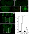
Fig. 1. Twsg1 gene expression and protein localization in the mandibular component of BA1
(A-F) Whole mount LacZ staining of heterozygous embryos carrying LacZ reporter gene in the Twsg1 locus. (A) Negative control at E9.5. (B) LacZ reporter gene is expressed throughout the mandibular arch at E9.5, in both distal and proximal regions. (C) Negative control at E10.5. (D) Gradient of the LacZ reporter gene expression at E10.5 with predominance in the distal region. (E) Negative control at E11.5. (F) The reporter gene is expressed in a pattern that is restricted toward the oral epithelium and proximal arch at E11.5. (G-O) TWSG1 protein localization by fluorescent immunostaining (right arch is shown). (G) DAPI, higher magnification of the image shown in M, (H) TWSG1 protein, (I) Overlay of DAPI and immunostaining for TWSG1 showing localization to the cell membranes, (J) DAPI, (K) Overlay of DAPI and TWSG1 immunostaining showing uniform distribution of TWSG1 throughout the mesenchyme of the mandibular arch at E9.5, (L) DAPI, (M) Overlay of DAPI and TWSG1 at E10.5 showing a gradient of distribution of TWSG1, (N) DAPI, (O) Overlay of DAPI and TWSG1 at E11.5 showing a shift in TWSG1 protein distribution toward the proximal BA1. Sections G-O are transverse sections; d, distal; p, proximal. Scale bar: 200 μm in A-F, 10 μm in G-I, 50 μm in J-O.
Fig. 2
Fig. 2. Histological analysis of BA1 derivatives at birth
(A) Wild type, hematoxylin and eosin staining of a sagittal section. (B,C) Wild type, coronal sections. (D) Twsg1−/− newborn, sagittal section. Absence of the mandible (m), anterior 2/3 of the tongue (t), underdeveloped nasal (n) and oral (o) cavities, hypoplastic pharynx (ph), lack of connection between the oral cavity and the pharynx in the mutants, but preservation of the hyoid bone (h) (BA2 derivative) in the mutants. (E,F) Twsg1−/− newborn, coronal sections. Absence of the mandible (m), tongue (t), upper incisors (ui), lower incisors (li), mandibular molars (mdm), and nasal septum (ns) in the mutants. Preservation of maxillary molars (mxm) in the mutants. Dashed lines with corresponding letters indicate the level of sectioning. Scale bar: 0.5 mm.
Fig. 3
Fig. 3. Ventral views of the whole-mount skull stained with alizarin red
(A) Wild-type ventral skull with normal mandibular bones (A') at birth. (B-D) Twsg1−/− skulls; (B) Ventral skull of Twsg1−/− newborn mouse with micrognathia showing deformity of the nasal capsule (NC), a longitudinal slit (LS) in the skull base, and preservation of the malleus (M) and pterygoid (PT). (B') Mandible in a micrognathic Twsg1−/− newborn mouse; the mandibular bones are fused in the midline indicated by an arrow. (C) Midline fusion of the right and left tympanic bones (T) in the agnathic Twsg1−/− mouse. (D) Loss or deformity of the majority of skeletal elements anterior to basioccipital (BO) with preservation of stapes (ST) in the Twsg1−/− mouse with anterior truncation phenotype. AL, alisphenoid; BS, basisphenoid; MX, maxilla; PL, palatine. Scale bar: 1 mm.
Fig. 4
Fig. 4. Spectrum of BA1 fusion inTwsg1−/− embryos at E9.5
(A,A') Wild type embryo in frontal and lateral views. (B,B'-D,D') Three classes of Twsg1 mutants. Class A - normal appearance of the BA1; Class B - partial midline fusion of BA1 with some preservation of the forebrain; Class C - severe midline fusion with ventral displacement of BA1 and severe reduction of anterior head structures. (E) Micro-dissected wild type BA1 at E10.5. (F) Mutant BA1 class C with severe midline fusion of the mandibular components and only partial separation of the maxillary components at E10.5. Dashed lines outline the mandibular prominences of BA1; mx, maxillary component of BA1; md, mandibular component of BA1. Scale bar: 200 μm.
Fig. 5
Fig. 5. Expression of distal markers in wild type and medially fused mutant BA1
(A,B) Lateral and frontal views of wild type Bmp4 expression in the distal region at E9.5. (C,D) Distal expression of Bmp4 is preserved in Twsg1 mutants but is shifted ventrally. (E,F) Lateral and frontal views of wild type Msx1 expression in the distal region at E9.5. (G,H) Distal expression of Msx1 is lost in the mutants at E9.5. (I,J) Gradient of wild type Msx2 expression in the distal region at E9.5; lateral and frontal views. (K,L) Distal expression of Msx2 is preserved in Twsg1 mutants and it is shifted ventrally. Note diffuse expression of Msx2 across BA1. (M) Wild type eHand expression in the distal region at E9.5. (N) Loss of distal expression of eHand in Twsg1−/− embryo at E9.5. (O) Wild type eHand expression in the distal region at E10.5. (P) Loss of distal expression of eHand in Twsg1−/− embryo at E10.5. Scale bar: 100 μm in A-D and 200 μm in E-P.
Fig. 6
Fig. 6. Expression of proximal markers in wild type and fused mutant BA1
(A,B) Lateral and frontal views of wild type Fgf8 expression in the oral epithelium of the proximal region of BA1 at E9.5. (C,D) The expression of Fgf8 is preserved in the proximal region of mutant embryos of class B and extends across the fused BA1 tissue, reflecting a shift of expression domain. (E,F) The expression of Fgf8 is significantly reduced in the proximal region of mutant embryos of class C, but present in the distal region. (G,H) Lateral and frontal views of wild type Barx1 expression in the mesenchyme of the proximal region of BA1 at E9.5. (I,J) Expression domain of Barx1 is extended across the midline in Twsg1 mutants. (K,L) Lateral and frontal views of wild type Nog expression in the oral epithelium of BA1 at E10.5. (I,J) Expression of Nog is extended across the fused BA1 in Twsg1−/− embryos. Scale bar: 200 μm in A-J and 100 μm in K-N.
Fig. 7
Fig. 7. Range of apoptosis in BA1 at E9.5
(A) Minimal apoptosis in wild type BA1. (B-E) Apoptosis in Twsg1−/− mutants. (B) Apoptosis in normal-appearing mutant BA1 is comparable to apoptosis in wild type BA1. (C) Increased apoptosis in the most distal part of mutant BA1. (D) More extensive apoptosis into the proximal region of mutant BA1. (E) Largely increased apoptosis throughout BA1 in the mutant. (F-H) Quantification of apoptosis. Comparison of apoptosis between wild type (F) and Twsg1 mutant (G) embryos at E9.5. The distal region was defined as the middle third of BA1. The area of each section was outlined within each box (dashed lines) and apoptotic cells were counted at 200x in each section per area (apoptotic index). (H) Data are expressed as the mean ± s.d. There was a significant 10-fold increase (*p<0.01) in apoptotic index in the distal region of the mutant vs. wild type BA1. Arrows point to mandibular prominences of BA1 and arrowheads point to the fused BA1. NS, not statistically significant, scale bar: 50 μm.
Fig. 8
Fig. 8. Distribution of pSMAD1/5 in BA1 at E9.5
(A-C) In wild type embryos, pSMAD1/5 immunostaining is distributed in a gradient, which is highest in the distal region and decreases proximally. (D, E) BMP signaling gradient is disrupted and shifted ventrally in Twsg1−/− embryos. (F) Negative control (no anti-pSMAD1/5 antibody). Few individual non-specifically staining cells can be seen. Scale bar: 50 μm.
Fig. 9
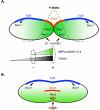
Fig. 9. Model of proximo-distal gene expression and BMP signaling in wild type andTwsg1 mutant BA1 at E9.5-E10.5
(A) Wild type. Bmp4 is expressed in the oral epithelium in the distal region and Fgf8 is expressed in the epithelium in the proximal region. Msx1 and Msx2 are downstream targets of BMP signaling in the distal region. Msx1 also receives input from ET- 1/eHAND. Gradient of BMP activity is indicated by green shading and a dashed line. Inhibition of BMP signaling by BMP-interacting proteins, for example TWSG1, would be more likely away from the site of synthesis where BMP4 level would be expected to be low (indicated by a dashed oval) to prevent further diffusion of BMP gradient. Alternatively, the role of BMP-binding proteins would be to help concentrate the ligand in the distal region to promote peak BMP activity in that region (indicated by a dotted oval). (B) Abnormally fused BA1 in Twsg1−/− embryo. Bmp4 as well as Msx2 expression is shifted ventrally. Loss of Msx1 expression may be secondary to loss of eHand expression. Fgf8 expression extends across the fused BA1. Ventral shift in Bmp4 expression is accompanied by disruption of BMP gradient and altered distribution of BMP signaling.

Similar articles

Cited by

References

    1. Ben-Zvi D, Shilo BZ, Fainsod A, Barkai N. Scaling of the BMP activation gradient in Xenopus embryos. Nature. 2008;453:1205–11. - PubMed
    1. Bollenbach T, Pantazis P, Kicheva A, Bokel C, Gonzalez-Gaitan M, Julicher F. Precision of the Dpp gradient. Development. 2008;135:1137–46. - PubMed
    1. Cerny R, Lwigale P, Ericsson R, Meulemans D, Epperlein HH, Bronner-Fraser M. Developmental origins and evolution of jaws: new interpretation of “maxillary” and “mandibular”. Dev Biol. 2004;276:225–36. - PubMed
    1. Chai Y, Jiang X, Ito Y, Bringas P, Jr., Han J, Rowitch DH, Soriano P, McMahon AP, Sucov HM. Fate of the mammalian cranial neural crest during tooth and mandibular morphogenesis. Development. 2000;127:1671–9. - PubMed
    1. Chai Y, Sasano Y, Bringas P, Jr., Mayo M, Kaartinen V, Heisterkamp N, Groffen J, Slavkin H, Shuler C. Characterization of the fate of midline epithelial cells during the fusion of mandibular prominences in vivo. Dev Dyn. 1997;208:526–35. - PubMed

Publication types

MeSH terms

LinkOut - more resources