. 2009 Apr 1;328(1):118-26.
doi: 10.1016/j.ydbio.2009.01.015. Epub 2009 Jan 20.

Jagged 1 is necessary for normal mouse lens formation

Affiliations

Jagged 1 is necessary for normal mouse lens formation

Tien T Le et al. Dev Biol. .

Abstract

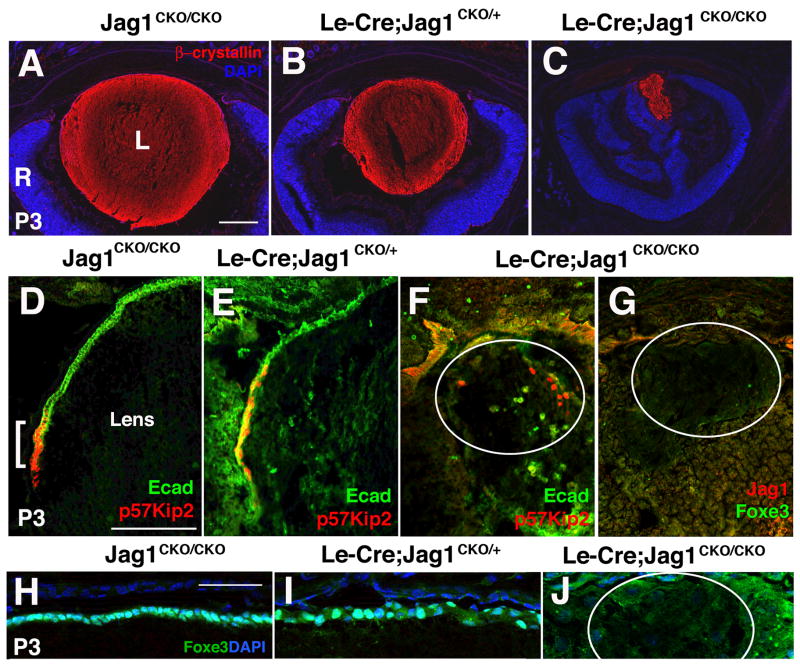
In mammals, two spatially and temporally distinct waves of fiber cell differentiation are crucial steps for normal lens development. In between these phases, an anterior growth zone forms in which progenitor cells migrate circumferentially, terminally exit the cell cycle and initiate differentiation at the lens equator. Much remains unknown about the molecular pathways orchestrating these processes. Previously, the Notch signal transduction pathway was shown to be critical for anterior lens progenitor cell growth and differentiation. However, the ligand or ligand(s) that direct these events are unknown. Using conditional gene targeting, we show that Jagged1 is required for lens fiber cell genesis, particularly that of secondary fiber cells. In the absence of Jagged1, the anterior growth and equatorial transition zones fail to develop fully, with only a handful of differentiated fiber cells present at birth. Adult Jagged1 conditional mutants completely lack lenses, along with severe anterior chamber deformities. Our data support the hypothesis that Jagged1-Notch signaling conveys a lateral inductive signal, which is indispensable for lens progenitor cell proliferation and differentiation.


Figures

Figure 1
Figure 1. Spatiotemporal expression of Jag1 mRNA and protein during mouse lens formation
In situ hybridization to detect Jag1 mRNA (A–E) and antibody labeling to visualize Jag1 protein (F–J) from E9.5 to birth. Jag1 is expressed in the lens placode beginning around E9.5 (arrows in A,F) and throughout the E10.5–11.5 lens vesicle (B,G). At these early ages Jag1 is also present in the distal optic vesicle and cup (arrowheads in A,B,F,G). Between E11.5-E12.5, Jag1 expression becomes localized to the posterior lens vesicle, where primary fiber cells differentiate (G,H). From E14.5 to at least P3, Jag1 mRNA and protein are predominantly expressed at the equatorial transition zone (D–E, I–J and data not shown). At these older ages, protein expression is broader than that of mRNA (compare E and J). Anterior is up in all panels; bar in A, C, F, H = 20 μm. OV = optic vesicle, OC = optic cup, LV = lens vesicle, L = lens, R = retina.
Figure 2
Figure 2. Adult phenotypes of Le-Cre;Jag1CKO/CKO eyes
A) Conditional deletion strategy to create Le-Cre; Jag1CKO/CKO lens mutants. B–D) Le-Cre; Jag1CKO/+ adult eyes are grossly indistinguishable from Jag1CKO/CKO littermate controls (n 3 per genotype). However, Le-Cre; Jag1CKO/CKO mutants have bilateral microphthalmia with missing fur and whiskers around the eye and snout (n>3). Mutant eyes lack pupillary openings (not shown). E–G) Histologic sections of these P21 eyes show slight reduction in the size of Le-Cre; Jag1CKO/+ lenses. Le-Cre; Jag1CKO/CKO eyes have essentially no lenses, although cellular debris resembling lens fiber cells could be found in some sections (arrows in G). Note complete absence of the anterior chamber and abnormally folded ciliary body/iris tissue that contains black pigment granules. Rostral is left in B–D; anterior up in E–G; bar = 500 μm in B; 5 μm in E,G. EE= ectoderm enhancer, P0 = Pax6 promoter, pA = poly A sequence, SS = signal sequence, DSL = Delta/Serrate/Lag domain, CR = cysteine-rich domain, TM = transmembrane domain, L = lens, R = retina, C = cornea, * = anterior chamber.
Figure 3
Figure 3. Postnatal Jag1 lens mutants have a profound loss of lens fibers and no AEL or transition zone
All panels contain sections of postnatal (P3) eyes. A–C) β-Crystallin expression in lens fiber cells (red) highlights the smaller size of Le-Cre; Jag1CKO/+lenses (B) and profound loss of this tissue in Le-Cre; Jag1CKO/CKO eyes (C). In the near absence of a lens, the retina (R, in blue) is abnormally folded. D–F) p57Kip2-E-cadherin (Ecad) double antibody labeling delineates the equatorial transition zone, where anterior progenitor cells (in green) exit mitosis (in red) to differentiate as secondary fiber cells. Normally there is minimal overlap of these two markers at the equator (bracket in D). In Le-Cre; Jag1CKO/+eyes, the p57Kip2+ domain is unusually elongated around the periphery, with inappropriate expression of both markers (E). However, in Le-Cre; Jag1CKO/CKO mutants, only sparse, randomly positioned, p57+ or Ecad+ cells were identifiable (white circle marks edges of lens tissue) (F). The p57Kip2+Ecad+ cells above the lens are in the ciliary body or cornea. G) Foxe3-Jag1 double antibody labeling confirms the loss of AEL and transition zone compartments, and removal of Jag1 protein from the lens in conditional mutants. F and G are sections nearby to that in C, note differences in magnification. H–J) AEL lens progenitor cell nuclei double labeled with anti-Foxe3 and DAPI. Normally these cells are tightly organized into a monolayer epithelium (H). But, Le-Cre; Jag1CKO/+ lenses the arrangement of Foxe3+ progenitor cell nuclei is abnormal. These cells are completely missing from the postnatal Jag1 mutant lens (circle in J). n=3 animals per genotype and marker. Anterior is up in all panels; bar in A–C = 5 μm, D–G = 50 μm, H–J = 100 μm. L = lens, R = retina.
Figure 4
Figure 4. Anterior epithelial lens cells critically require Jag1
A–F) E12.5 lens sections double labeled with Foxe3 (green) and Ecad (red). The expression of both markers is restricted to lens progenitor cells in the anterior epithelial layer (AEL). In Le-Cre;Jag1CKO/+ and Le-Cre;Jag1CKO/CKO eyes, there is a progressive loss of both Foxe3 and Ecad in the anterior lens (arrows in all panels, n = 4 embryos/genotype). Note Ecad expression in the forming cornea appears unaffected (arrowheads in D–F, J–L). (G–L) At E14.5, the smaller size of Le-Cre;Jag1CKO/+ and Le-Cre;Jag1CKO/CKO lenses is apparent, along with dramatic reduction of Foxe3 and Ecad expression. For each marker, imaging exposure time was held constant among genotypes at each age. Among four E14.5 Jag1 lens mutant embryos, half had no anterior Foxe3+ cells (arrow in I), and the rest a handful of Foxe3+ AEL cells (not shown). Insets in panels F and L show that DAPI-labeled fiber cell nuclei are appropriately positioned in mutant lenses. Low level, patchy fluorescence in the fiber cell compartment sometimes occurred (H,I), but was not meaningful since control sections lacking primary antibody exhibit the same pattern. Ecad expression in the AEL (arrows) is abnormal in both K and L. Anterior is up in all panels; bar = 20 μm. L = lens, C = cornea.
Figure 5
Figure 5. Loss of Jag1 affects both lens progenitor cell proliferation and differentiation
A) The percentage of fiber cells (Foxe3-negative/total cells) is increased at E10.5 and E14.5 Le-Cre;Jag1CKO/CKO lenses. B–L) Progenitor cell cycle markers in Jag1 lens mutants. B) BrdU+ cells are significantly decreased at all ages analyzed. C) Cyclin D1+ cells are lost in E10.5 and E14.5 Le-Cre;Jag1CKO/CKO lenses, but rebound above normal levels at E12.5. D) By contrast, Cyclin D2+ cells are not significantly unaffected. E) There are significantly fewer p27Kip1+ cells during secondary fiber cell genesis in E12.5-E14.5 Le-Cre;Jag1CKO/CKO eyes. F) The percentage of p57Kip2+ cells correlates with increased fiber cells only at E14.5. G–L) Example Foxe3/DAPI (G–I) BrdU/Ecad (J–L) and CyclinD1/Ecad (M–O) labeled lenses used for quantification in A–C (DAPI channel omitted in J–O) showing obvious reduction of BrdU+ and Cyclin D1+ nuclei in I and L respectively. Note the split Ecad expression domain in L, with both CyclinD1 and Ecad expression missing from the central AEL. Bar graphs show mean ± s.e.m., *p < 0.05, **p < 0.01, ***p < 0.001. LP = lens pit; L = lens; bar in panels G, M = 20 μm.
Figure 6
Figure 6. Jag1-Notch signaling in the lens is not bidirectional
A–C) As early as E10.5, Jag1 protein is reduced in Le-Cre;Jag1CKO/+ and total loss in Le-Cre;Jag1CKO/CKO lens pits (arrowheads point to Jag1+ cell membranes in white; n = 3 embryos/genotype). However, Jag1 in the distal optic cup (denoted by asterisks) is unaffected. D–E) The Notch pathway effector Hes1 (green) is not obviously affected in E10.5 Le-Cre;Jag1CKO/+or Le-Cre;Jag1CKO/CKO mutants (arrows point to Hes1+ nuclei, arrowheads point to Jag1 in cell membranes). G–I) By E12.5, Hes1 is totally missing from Jag1 mutant lenses (arrows in G–I, n = 3 embryos/genotype), two days earlier than in Le-Cre;RbpjCKO/CKO (Figure 1G of Rowan et al, 2008). Fuchsia brackets denote unaffected Hes1 expression in the hyaloid vasculature immediately posterior to the lens. J–L) Importantly, Jag1 expression appears normal in Le-Cre;RbpjCKO/CKO lenses (compare K to J; n = 3 embryos/genotype). GFP expression in the inset in I and in panel L indicates cells with Cre activity. LP = lens pit; L = lens; Bar in A, G = 20 μm.

Similar articles

Cited by

References

    1. Ashery-Padan R, Marquardt T, Zhou X, Gruss P. Pax6 activity in the lens primordium is required for lens formation and for correct placement of a single retina in the eye. Genes Dev. 2000;14:2701–11. - PMC - PubMed
    1. Bao ZZ, Cepko CL. The expression and function of Notch pathway genes in the developing rat eye. Journal of Neuroscience. 1997;17:1425–34. - PMC - PubMed
    1. Beebe D, Garcia C, Wang X, Rajagopal R, Feldmeier M, Kim JY, Chytil A, Moses H, Ashery-Padan R, Rauchman M. Contributions by members of the TGFbeta superfamily to lens development. Int J Dev Biol. 2004;48:845–56. - PubMed
    1. Blixt A, Landgren H, Johansson BR, Carlsson P. Foxe3 is required for morphogenesis and differentiation of the anterior segment of the eye and is sensitive to Pax6 gene dosage. Dev Biol. 2007;302:218–29. - PubMed
    1. Blixt A, Mahlapuu M, Aitola M, Pelto-Huikko M, Enerback S, Carlsson P. A forkhead gene, FoxE3, is essential for lens epithelial proliferation and closure of the lens vesicle. Genes Dev. 2000;14:245–54. - PMC - PubMed

Publication types

MeSH terms

LinkOut - more resources