

Magnetic nanoparticles for theragnostics
- PMID: 19389434
- PMCID: PMC2700776
- DOI: 10.1016/j.addr.2009.03.007
Magnetic nanoparticles for theragnostics
Abstract
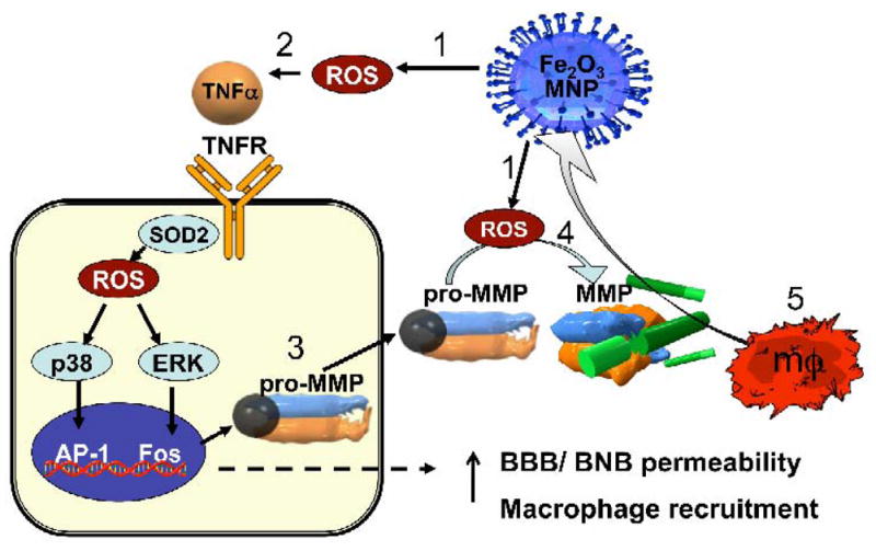
Engineered magnetic nanoparticles (MNPs) represent a cutting-edge tool in medicine because they can be simultaneously functionalized and guided by a magnetic field. Use of MNPs has advanced magnetic resonance imaging (MRI), guided drug and gene delivery, magnetic hyperthermia cancer therapy, tissue engineering, cell tracking and bioseparation. Integrative therapeutic and diagnostic (i.e., theragnostic) applications have emerged with MNP use, such as MRI-guided cell replacement therapy or MRI-based imaging of cancer-specific gene delivery. However, mounting evidence suggests that certain properties of nanoparticles (e.g., enhanced reactive area, ability to cross cell and tissue barriers, resistance to biodegradation) amplify their cytotoxic potential relative to molecular or bulk counterparts. Oxidative stress, a 3-tier paradigm of nanotoxicity, manifests in activation of reactive oxygen species (ROS) (tier I), followed by a proinflammatory response (tier II) and DNA damage leading to cellular apoptosis and mutagenesis (tier III). Invivo administered MNPs are quickly challenged by macrophages of the reticuloendothelial system (RES), resulting in not only neutralization of potential MNP toxicity but also reduced circulation time necessary for MNP efficacy. We discuss the role of MNP size, composition and surface chemistry in their intracellular uptake, biodistribution, macrophage recognition and cytotoxicity, and review current studies on MNP toxicity, caveats of nanotoxicity assessments and engineering strategies to optimize MNPs for biomedical use.
Figures

References
-
- Whitesides GM. The ‘right’ size in nanobiotechnology. Nat Biotechnol. 2003;21(10):1161–5. - PubMed
-
- Gupta AK, Gupta M. Synthesis and surface engineering of iron oxide nanoparticles for biomedical applications. Biomaterials. 2005;26(18):3995–4021. - PubMed
-
- Gupta AK, et al. Recent advances on surface engineering of magnetic iron oxide nanoparticles and their biomedical applications. Nanomed. 2007;2(1):23–39. - PubMed
-
- McCarthy JR, et al. Targeted delivery of multifunctional magnetic nanoparticles. Nanomed. 2007;2(2):153–67. - PubMed
-
- Duguet E, et al. Magnetic nanoparticles and their applications in medicine. Nanomed. 2006;1(2):157–68. - PubMed
Publication types
MeSH terms
Substances
Grants and funding
LinkOut - more resources
Full Text Sources
Other Literature Sources
