. 2009 Apr 28;53(17):1517-27.
doi: 10.1016/j.jacc.2008.12.056.

Thin-walled microvessels in human coronary atherosclerotic plaques show incomplete endothelial junctions relevance of compromised structural integrity for intraplaque microvascular leakage

Affiliations

Thin-walled microvessels in human coronary atherosclerotic plaques show incomplete endothelial junctions relevance of compromised structural integrity for intraplaque microvascular leakage

Judith C Sluimer et al. J Am Coll Cardiol. .

Abstract

Objectives: This study sought to examine the ultrastructure of microvessels in normal and atherosclerotic coronary arteries and its association with plaque phenotype.

Background: Microvessels in atherosclerotic plaques are an entry point for inflammatory and red blood cells; yet, there are limited data on the ultrastructural integrity of microvessels in human atherosclerosis.

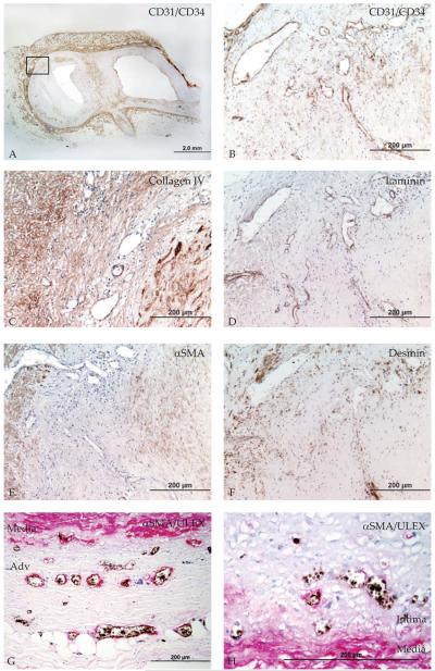
Methods: Microvessel density (MVD) and ultrastructural morphology were determined in the adventitia, intima-media border, and atherosclerotic plaque of 28 coronary arteries using immunohistochemistry for endothelial cells (Ulex europeaus, CD31/CD34), basement membrane (laminin, collagen IV), and mural cells (desmin, alpha-smooth muscle [SM] actin, smoothelin, SM1, SM2, SMemb). Ultrastructural characterization of microvessel morphology was performed by electron microscopy.

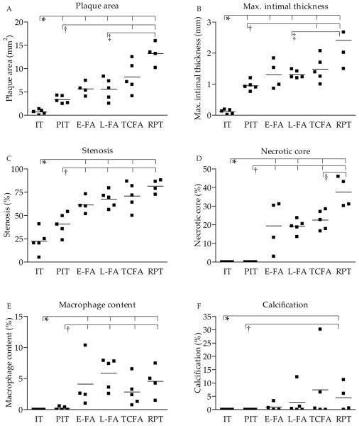
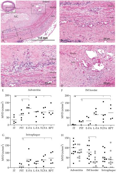
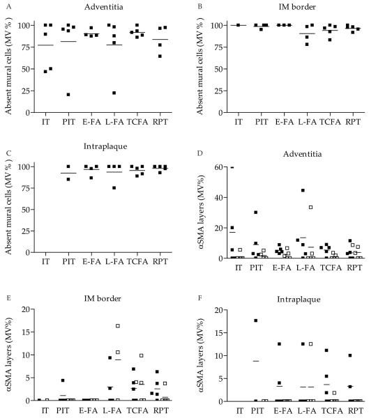
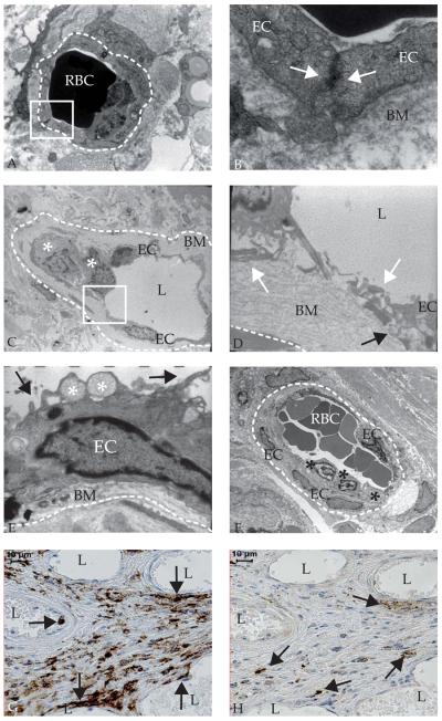
Results: The MVD was increased in advanced plaques compared with early plaques, which correlated with lesion morphology. Adventitial MVD was higher than intraplaque MVD in normal arteries and early plaques, but adventitial and intraplaque MVD were similar in advanced plaques. Although microvessel basement membranes were intact, the percentage of thin-walled microvessels was similarly low in normal and atherosclerotic adventitia, in the adventitia and the plaque, and in all plaque types. Intraplaque microvascular endothelial cells (ECs) were abnormal, with membrane blebs, intracytoplasmic vacuoles, open EC-EC junctions, and basement membrane detachment. Leukocyte infiltration was frequently observed by electron microscopy, and confirmed by CD45RO and CD68 immunohistochemistry.

Conclusions: The MVD was associated with coronary plaque progression and morphology. Microvessels were thin-walled in normal and atherosclerotic arteries, and the compromised structural integrity of microvascular endothelium may explain the microvascular leakage responsible for intraplaque hemorrhage in advanced human coronary atherosclerosis.


Figures

Figure 1
Figure 1. Atherosclerotic plaque morphology in normal and atherosclerotic coronary arteries
A. Plaque area, B. maximal intimal thickness, C. stenosis, D. necrotic core content, E. macrophage content, and F. calcification content in normal coronary artery and various coronary plaque types. * p-value<0.05 vs. IT; † p<0.05 vs. PIT; ‡ p<0.05 vs. L-FA; § p<0.05 vs. TCFA. Abbreviations: IT, normal intimal thickening; PIT, pathological intimal thickening; FA, thick fibrous cap atheroma with an early or (E-FA) late necrotic core (L-FA); TCFA, thin fibrous cap atheroma; RPT, ruptured plaque
Figure 2
Figure 2. Microvessel density increases with atherosclerotic progression
A. Detail of hematoxylin and eosin stained coronary artery with thick cap fibroatheroma (NC, necrotic core), inset shows entire coronary artery. Boxed regions B-D illustrate the regions of interest and correspond to photographs 2B showing microvessels in the adventitia, 2C intima-media border, and D. in the intraplaque region. E. Mean microvessel density (MVD) was quantified in adventitia, F. in the IM border, and G. in the intraplaque region, and increased with progression. H. Microvessel density was higher in concentric (black squares) than in eccentric (open squares) plaques. * p-value<0.05 vs. IT; † p<0.05 vs. PIT; ‡ p<0.05 eccentric vs. concentric; Abbreviations: IT, normal intimal thickening; PIT, pathological intimal thickening; FA, thick fibrous cap atheroma with an early or (E-FA) late necrotic core (L-FA); TCFA, thin fibrous cap atheroma; RPT, ruptured plaque
Figure 3
Figure 3. Basement membrane and mural cell coverage in normal and atherosclerotic coronary arteries
A. Coronary fibroatheroma stained with CD31/CD34 cocktail. Boxed region indicates origin of B. CD31+CD34+ microvessels run from the adventitia through the media into the plaque. C Adjacent section stained with laminin or D. collagen IV, showing intact microvessel basement membrane in the adventitia, intima-media (IM) border and plaque. E. Adjacent section stained with alpha smooth muscle actin (aSMA), showing mostly αSMA microvessels in the adventitia, IM border and plaque. F. Adjacent section stained with desmin shows some desmin+ microvessels in the adventitia, IM border and intraplaque region. G Adjacent section double-stained with lectin-1 (black) and αSMA (red) shows infrequent mural cell coverage in the adventitia and H. in the IM border.
Figure 4
Figure 4. Quantification of mural cell coverage in various plaque types
A. Mural cell coverage was low in the adventitia, B. intima-media (IM) border, and C. intraplaque region of both normal and atherosclerotic coronary arteries. D. Percentage of microvessels with single (black squares) and multiple (open squares) mural cell layers in adventitia, E. IM border and F. intraplaque region. All p-values>0.05. Abbreviations: IT, normal intimal thickening; PIT, pathological intimal thickening; FA, thick fibrous cap atheroma with an early or (E-FA) late necrotic core (L-FA); TCFA, thin fibrous cap atheroma; RPT, ruptured plaque
Figure 5
Figure 5. Intraplaque microvessels show abnormal endothelial cell (EC) morphology, aberrant junctions, and leukocyte infiltration as demonstrated by electron microscopy
A. Ultrastructure of an adventitial microvessel (dashed line indicates circumference) in a non-diseased coronary artery with luminal red blood cells (RBC) (electron microscopy (EM, 8000x) B. Magnification of the boxed region in A of microvessel with intact basement membrane (BM) and inter-endothelial junction indicated by close contact (white arrows) between endothelial cells (20,000x) C. Ultrastructure of intraplaque microvessel with leukocytes (white asterisks) (650x) D. Magnification of the boxed region in C, showing aberrant inter-EC junction (white arrows) and basement membrane detachment (black arrow) (4600x) E. Dysfunctional EC ultrastructure in an intraplaque microvessel (6300x): membrane blebs (black arrow) and intracytoplasmic vacuoles (white asterisk) F. Leukocytes (asterisks) adhering to intraplaque microvessel endothelium (460x) G. Immunohistochemistry shows CD45+ cells in and near microvessels (arrows). H. Immunohistochemistry showing mast cell tryptase positive cells at larger distance from microvessels (arrows). L, lumen.

Comment in

References

    1. Moreno PR, Purushothaman KR, Fuster V, et al. Plaque neovascularization is increased in ruptured atherosclerotic lesions of human aorta: implications for plaque vulnerability. Circulation. 2004;110:2032–8. - PubMed
    1. Virmani R, Kolodgie FD, Burke AP, et al. Atherosclerotic Plaque Progression and Vulnerability to Rupture Angiogenesis as a Source of Intraplaque Hemorrhage. Arterioscler Thromb Vasc Biol. 2005;25:2054–61. - PubMed
    1. Bot I, de Jager SC, Zernecke A, et al. Perivascular mast cells promote atherogenesis and induce plaque destabilization in apolipoprotein E-deficient mice. Circulation. 2007;115:2516–25. - PubMed
    1. Kockx MM, Cromheeke KM, Knaapen MW, et al. Phagocytosis and macrophage activation associated with hemorrhagic microvessels in human atherosclerosis. Arterioscler Thromb Vasc Biol. 2003;23:440–6. - PubMed
    1. Kolodgie FD, Gold HK, Burke AP, et al. Intraplaque hemorrhage and progression of coronary atheroma. N Engl J Med. 2003;349:2316–25. - PubMed

Publication types