

Combination of reverse and chemical genetic screens reveals angiogenesis inhibitors and targets
- PMID: 19389629
- PMCID: PMC3984492
- DOI: 10.1016/j.chembiol.2009.02.010
Combination of reverse and chemical genetic screens reveals angiogenesis inhibitors and targets
Abstract
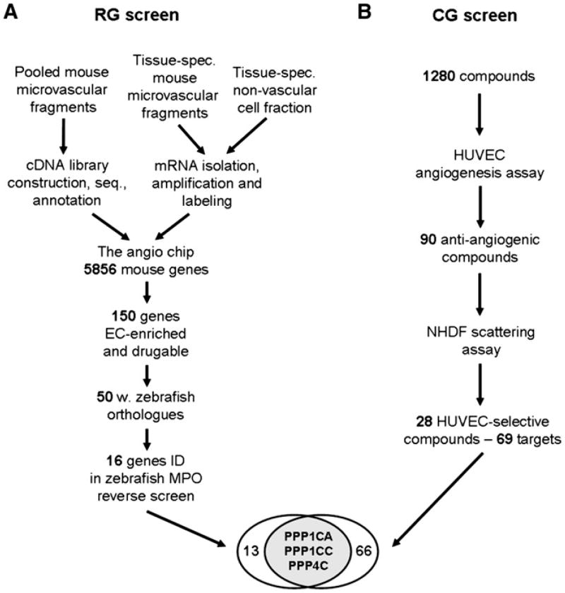
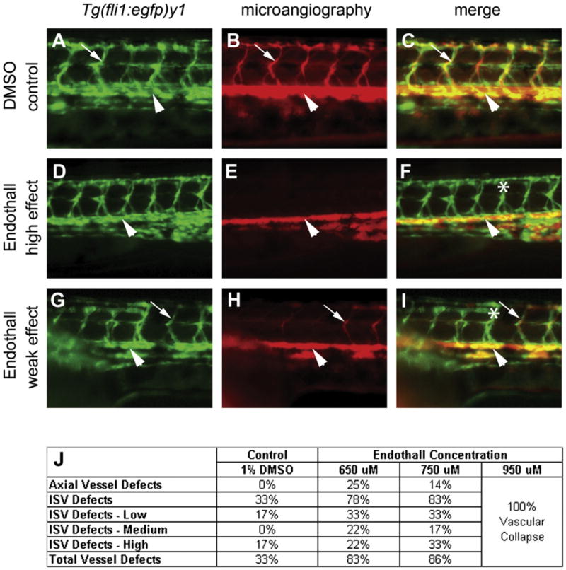
We combined reverse and chemical genetics to identify targets and compounds modulating blood vessel development. Through transcript profiling in mice, we identified 150 potentially druggable microvessel-enriched gene products. Orthologs of 50 of these were knocked down in a reverse genetic screen in zebrafish, demonstrating that 16 were necessary for developmental angiogenesis. In parallel, 1280 pharmacologically active compounds were screened in a human cell-based assay, identifying 28 compounds selectively inhibiting endothelial sprouting. Several links were revealed between the results of the reverse and chemical genetic screens, including the serine/threonine (S/T) phosphatases ppp1ca, ppp1cc, and ppp4c and an inhibitor of this gene family; Endothall. Our results suggest that the combination of reverse and chemical genetic screens, in vertebrates, is an efficient strategy for the identification of drug targets and compounds that modulate complex biological systems, such as angiogenesis.
Figures
Similar articles
-
Identification of novel angiogenesis inhibitors.Bioorg Med Chem Lett. 2005 Sep 15;15(18):4125-9. doi: 10.1016/j.bmcl.2005.06.001. Bioorg Med Chem Lett. 2005. PMID: 15993586
-
A zebrafish in vivo phenotypic assay to identify 3-aminothiophene-2-carboxylic acid-based angiogenesis inhibitors.Assay Drug Dev Technol. 2014 Nov-Dec;12(9-10):527-35. doi: 10.1089/adt.2014.606. Assay Drug Dev Technol. 2014. PMID: 25506802 Free PMC article.
-
Eriocalyxin B, a natural diterpenoid, inhibited VEGF-induced angiogenesis and diminished angiogenesis-dependent breast tumor growth by suppressing VEGFR-2 signaling.Oncotarget. 2016 Dec 13;7(50):82820-82835. doi: 10.18632/oncotarget.12652. Oncotarget. 2016. PMID: 27756875 Free PMC article.
-
Antiangiogenic cancer drug using the zebrafish model.Arterioscler Thromb Vasc Biol. 2014 Sep;34(9):1846-53. doi: 10.1161/ATVBAHA.114.303221. Epub 2014 Jun 5. Arterioscler Thromb Vasc Biol. 2014. PMID: 24903092 Review.
-
A critical analysis of current in vitro and in vivo angiogenesis assays.Int J Exp Pathol. 2009 Jun;90(3):195-221. doi: 10.1111/j.1365-2613.2008.00633.x. Int J Exp Pathol. 2009. PMID: 19563606 Free PMC article. Review.
Cited by
-
RACK1 contributes to the upregulation of embryonic genes in a model of cardiac hypertrophy.Sci Rep. 2024 Oct 28;14(1):25698. doi: 10.1038/s41598-024-76138-x. Sci Rep. 2024. PMID: 39465301 Free PMC article.
-
Female mice lacking Pald1 exhibit endothelial cell apoptosis and emphysema.Sci Rep. 2017 Nov 13;7(1):15453. doi: 10.1038/s41598-017-14894-9. Sci Rep. 2017. PMID: 29133847 Free PMC article.
-
Generation of Rab-based transgenic lines for in vivo studies of endosome biology in zebrafish.Dev Dyn. 2011 Nov;240(11):2452-65. doi: 10.1002/dvdy.22758. Epub 2011 Oct 4. Dev Dyn. 2011. PMID: 21976318 Free PMC article.
-
Zebrafish xenotransplantation as a tool for in vivo cancer study.Fam Cancer. 2015 Sep;14(3):487-93. doi: 10.1007/s10689-015-9802-3. Fam Cancer. 2015. PMID: 25860646 Review.
-
Phenotype-based high-content chemical library screening identifies statins as inhibitors of in vivo lymphangiogenesis.Proc Natl Acad Sci U S A. 2012 Oct 2;109(40):E2665-74. doi: 10.1073/pnas.1206036109. Epub 2012 Sep 4. Proc Natl Acad Sci U S A. 2012. PMID: 22949700 Free PMC article.
References
-
- Adams RH, Alitalo K. Molecular regulation of angiogenesis and lymphangiogenesis. Nat Rev Mol Cell Biol. 2007;8:464–478. - PubMed
-
- Andreoli CM, Miller JW. Anti-vascular endothelial growth factor therapy for ocular neovascular disease. Curr Opin Ophthalmol. 2007;18:502–508. - PubMed
-
- Benjamini Y, Hochberg Y. Controlling the false discovery rate: a practical and powerful approach to multiple testing. J Royal Stat Soc B (Methodological) 1995;57:289–300.
-
- Bondjers C, He L, Takemoto M, Norlin J, Asker N, Hellstrom M, Lindahl P, Betsholtz C. Microarray analysis of blood microvessels from PDGF-B and PDGF-Rβ mutant mice identifies novel markers for brain pericytes . FASEB J. 2006;20:1703–1705. - PubMed
MeSH terms
Substances
Associated data
- Actions
Grants and funding
LinkOut - more resources
Full Text Sources
Molecular Biology Databases
