. 2009 Jul 10;284(28):18851-62.
doi: 10.1074/jbc.M808605200. Epub 2009 Apr 23.

Target gene specificity of USF-1 is directed via p38-mediated phosphorylation-dependent acetylation

Affiliations

Target gene specificity of USF-1 is directed via p38-mediated phosphorylation-dependent acetylation

Sébastien Corre et al. J Biol Chem. .

Abstract

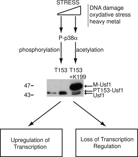
How transcription factors interpret the output from signal transduction pathways to drive distinct programs of gene expression is a key issue that underpins development and disease. The ubiquitously expressed basic-helix-loop-helix leucine zipper upstream stimulating factor-1 binds E-box regulatory elements (CANNTG) to regulate a wide number of gene networks. In particular, USF-1 is a key component of the tanning process. Following UV irradiation, USF-1 is phosphorylated by the p38 stress-activated kinase on threonine 153 and directly up-regulates expression of the POMC, MC1R, TYR, TYRP-1 and DCT genes. However, how phosphorylation on Thr-153 might affect the activity of USF-1 is unclear. Here we show that, in response to DNA damage, oxidative stress and cellular infection USF-1 is acetylated in a phospho-Thr-153-dependent fashion. Phospho-acetylated USF-1 is nuclear and interacts with DNA but displays altered gene regulatory properties. Phospho-acetylated USF-1 is thus proposed to be associated with loss of transcriptional activation properties toward several target genes implicated in pigmentation process and cell cycle regulation. The identification of this critical stress-dependent USF-1 modification gives new insights into understanding USF-1 gene expression modulation associated with cancer development.


Figures

FIGURE 1.
FIGURE 1.
Identification of a new USF-1 stress-responsive form. Western blot analysis of 501mel cell extracts after cells treatment using anti-USF-1, anti-USF-2, anti-P-p38, and anti-β-tubulin or anti-Lamin B antibodies as loading control. A, cells recovered 1.5 h after UVB irradiation (312 nm 80 J/m2), menadione (0.1 μm), and MMS (0.2 mm). B, 501mel cells extracts treated for 24 h with DNA-damaging compounds (hydroxyurea (HA): 2 μm, methyl methane sulfonate (MMS): 0.2 mm) and heavy metal compounds (cadmium (Cd): 5 μm; arsenic (As): 5 μm). C, 501mel cells treated for 4 h with increasing concentrations of hydrogen peroxide (from 0 to 10 mm). D, 501mel cells stimulated for 24 h using critical H2O2 concentrations (10, 40, and 100 μm) in the presence or not of the specific p38 kinase inhibitor (SB203580 10 μm, 1 h before induction). E, the impact of oxidative stress on cell viability is assessed using an 3-(4,5-dimethylthiazol-2-yl)-2,5-diphenyltetrazolium bromide test after 24-h H2O2 treatment (from 50 nm to 100 mm). F, M. hyorrhinis contamination (black arrow) of BLM1492 cells: contamination level is monitored by immunofluorescence using DNA staining (Hoechst) (negative image) and a time course induction of M-USF-1 formation is visualized by Western blotting analysis in relation to contamination level. G, CS6269 cells were infected with vesicular stomatitis virus (VSV) using gradual dilutions (1:250, 1:500, and 1:1000) of a virus stock titrated at 5.106 plaque forming units per ml (pfu/ml). 24-h post-infection cells were recovered for Western blotting analysis.
FIGURE 2.
FIGURE 2.
Implication of p38 pathway in M-USF-1 form and identification. Western blot analysis of 501mel cell extracts after cells treatment using anti-USF-1, anti-USF-2, anti-P-p38, and anti-β-tubulin or anti-Lamin B antibodies as loading control. A, cells recovered 24 h post-stress stimulation (MMS, 0.2 mm), in the presence or not of specific kinase inhibitors, added to the cell culture 1 h prior to the MMS stimulation. B, pretreated 501mel cells with the p38-specific kinase inhibitor (SB 203580, 10 μm) 1 h prior to UVB (80 J/m2) stimulations. C, purification of the phosphoprotein fraction of mycoplasma-infected BLM1492 cells using the PhosphoProtein purification kit (Qiagen®). Aliquots of total cell extract, wash 1 and 2 (W1 and W2) and purified phosphoprotein (PP) fractions were resolved on SDS-PAGE and analyzed by Western blotting analysis using anti-USF-1 Ab (Santa Cruz Biotechnology), p38 Ab (Cell Signaling), and P-p38 Ab (Cell Signaling). D, immunoprecipitations of USF-1 were performed on total cell extract from 501mel cells highly contaminated with mycoplasma, using specific anti-USF-1 Ab. In these cells, M-USF-1 protein is mainly present and thus facilitates the identification of post-translational modification of M-USF-1 by Western blot analysis. We load into the gel two different concentrations of recovered protein (high: 1, low: 2) to distinguish the different post-translational modifications of USF-1. E, Western blot analysis of Mycoplasma-infected BLM1492 cells, obtained 12 h after stimulation in the presence of different concentrations the specific deacetylases inhibitor trichostatin A (0–10 μm).
FIGURE 3.
FIGURE 3.
Modification of M-USF-1 corresponds to acetylation on Lysine 199. To identify the potential position of the M-USF-1 modifications, lysine residues were mutated to arginine. A, structure and position of the mutated lysines. B, wild type (WT) and mutated (T153E, T153A, K188R, R199R, K214R, K286R, K288R, and K306R) recombinant USF-1 proteins transiently expressed in 501mel cells, followed by UVB induction (400 J/m2) or not, were resolved on an SDS-PAGE and analyzed using SV5-tagged antibody. Previous to the induction, sodium butyrate (deacetylase inhibitor, 10 mm final) was added to the culture medium. Anti-Lamin B was used as loading control. C, immunoprecipitation of WT, T153A, and K199R recombinant proteins expressed in BLM 1492 mycoplasma-positive cell line using anti-USF-1 antibody. The presence of post-transcriptional modifications in response to stress stimuli (mycoplasma infection) was analyzed by Western blot using anti-SV5, and anti-acetylated K antibodies.
FIGURE 4.
FIGURE 4.
M-USF-1 interacts in vivo and in vitro with DNA. A, Western blot analysis (anti-USF-1 antibody) of fractionated cell extract, obtained 12 h after stress stimulation (MMS, 0. 2 mm). B, Western blot analysis is performed on ChIP extract to confirm that M-USF-1 was exclusively present in MMS-treated cells. C, schematic representation of human POMC, MC1R, TYR, CCNB1, CDC2, TERT, and HSP70 promoters. E-box motifs are shown as gray rectangles. Positions of PCR primers (→←) used for chromatin immunoprecipitation assays are shown relative to the start sites of each promoter, and are centered on each E-box motif, except for HSP70 primers. D, ChIP assays are performed on 501mel cells, which were stimulated or not with MMS (0.2 mm for 24 h), using anti-USF-1 antibody or nonspecific IgG. Recovered DNA was subjected to PCR using specific primers of each promoter. Specific primers for the Hsp70 promoter gene were used as negative control. E, DNA-binding mobility assay were performed in the presence of mycoplasma-free (Ctr) and contaminated BLM 1492 whole cell extracts and following transfection of different USF-1 mutants in 501mel cells, using both concensus Mbox and derivated Ebox for TYR promoter-labeled probes. USF-1 is shifted with specific anti-USF-1 or anti-SV5 FLAG antibodies, and competition is performed with cold probes.
FIGURE 5.
FIGURE 5.
Transcriptional activity of the M-USF-1 form. Expression level of USF-1 target genes were analyzed under specific stress conditions, using quantitative reverse transcription-PCR. A, gene expression levels of USF-1 target genes (pigmentation genes: POMC, MC1R, and TYR and cell cycle genes: CCNB1, CDC2, and TERT) were analyzed by real-time PCR over a single time course (12 h) using MMS-treated (0.2 mm) 501mel cells. USF-1 expression level, which remains constant over the 12-h induction, was used as the calibrator gene and the non-induced condition as the reference. B, Western blot analysis of USF-1 phosphorylation state (anti-USF-1 Ab, Santa Cruz Biotechnology), was performed on protein samples prepared from 501mel cells, respectively, over the 12-h time course after MMS stimulation. C, luciferase assays with PGL3-TYR promoter (−300/+80) in the presence of different constructs for USF-1 after or not treatment by different doses of H2O2 (200 μm and 1 mm), which, respectively, induces activation of USF-1 by phosphorylation of Thr-153 and apparition of M-USF-1, confirmed by Western blot analysis of 501mel cells extracts 48 h after cotransfection, were performed in 501mel cells to analyze the transcriptional activity of USF-1 after modification.
FIGURE 6.
FIGURE 6.
Phospho-dependant acetylation of USF-1 is associated with a decrease of transcriptional activity. A, luciferase assays with PGL3-TYR promoter (−300/+80) after or not treatment by different doses of H2O2 (200 μm and 1 mm), which, respectively, induces activation of USF-1 by phosphorylation of Thr-153 and apparition of M-USF-1 in the presence of different doses of trichostatin A, confirmed by Western blot analysis of 501mel cells extracts 48 h after cotransfection. B, luciferase assays with PGL3-TYR promoter (−300/+80) in the presence of different forms of USF-1 after or not treatment by different doses of H2O2 and trichostatin A. Phosphorylation and acetylation is confirmed by Western blot analysis.
FIGURE 7.
FIGURE 7.
USF-1 gene expression regulation model. Following, low levels of stress, the USF-1 transcription factor is activated through phosphorylation of the Thr-153 residue (PT153-USF-1) in a p38-dependent manner, leading to the up-regulation of USF target gene networks allowing the tanning response, cell cycle, and proliferation control. When stress is above a critical threshold (STRESS), the USF-1 transcription factor is acetylated on Lys-199 in a phospho-Thr-153-dependent fashion, leading to the appearance of a 47-kDa form (M-Usf1), associated with loss of transcriptional activation.

Similar articles

Cited by

References

    1. Corre S., Galibert M. D. (2005) Pigment Cell Res. 18, 337–348 - PubMed
    1. Sirito M., Walker S., Lin Q., Kozlowski M. T., Klein W. H., Sawadogo M. (1992) Gene Expr. 2, 231–240 - PMC - PubMed
    1. Sirito M., Lin Q., Maity T., Sawadogo M. (1994) Nucleic Acids Res. 22, 427–433 - PMC - PubMed
    1. Sirito M., Lin Q., Deng J. M., Behringer R. R., Sawadogo M. (1998) Proc. Natl. Acad. Sci. U.S.A. 95, 3758–3763 - PMC - PubMed
    1. Casado M., Vallet V. S., Kahn A., Vaulont S. (1999) J. Biol. Chem. 274, 2009–2013 - PubMed

Publication types