. 2009 Jul 10;284(28):18699-706.
doi: 10.1074/jbc.M109.005819. Epub 2009 Apr 22.

{alpha}MSH and Cyclic AMP elevating agents control melanosome pH through a protein kinase A-independent mechanism

Affiliations

{alpha}MSH and Cyclic AMP elevating agents control melanosome pH through a protein kinase A-independent mechanism

Yann Cheli et al. J Biol Chem. .

Abstract

Melanins are synthesized in melanocytes within specialized organelles called melanosomes. Numerous studies have shown that the pH of melanosome plays a key role in the regulation of melanin synthesis. However, until now, acute regulation of melanosome pH by a physiological stimulus has never been demonstrated. In the present study, we show that the activation of the cAMP pathway by alphaMSH or forskolin leads to an alkalinization of melanosomes and a concomitant regulation of vacuolar ATPases and ion transporters of the solute carrier family. The solute carrier family members include SLC45A2, which is mutated in oculocutaneous albinism type IV, SLC24A4 and SLC24A5, proteins implicated in the control of eye, hair, and skin pigmentation, and the P protein, encoded by the oculocutaneous albinism type II locus. Interestingly, H89, a pharmacological inhibitor of protein kinase A (PKA), prevents the cAMP-induced pigmentation and induces acidification of melanosomes. The drastic depigmenting effect of H89 is not due to an inhibition of tyrosinase expression. Indeed, H89 blocks the induction of melanogenesis induced by LY294002, a potent inhibitor of the PI 3-kinase pathway, without any effect on tyrosinase expression. Furthermore, PKA is not involved in the inhibition of pigmentation promoted by H89 because LY294002 induces pigmentation independently of PKA. Also, other PKA inhibitors do not affect pigmentation. Taken together, our results strengthen the support for a key role of melanosome pH in the regulation of melanin synthesis and, for the first time, demonstrate that melanosome pH is regulated by cAMP and alphaMSH. Notably, these are both mediators of the response to solar UV radiation, the main physiological stimulus of skin pigmentation.


Figures

FIGURE 1.
FIGURE 1.
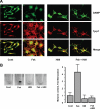
Alkalinization of melanosomes by αMSH and forskolin. B16 mouse melanoma cells were incubated with or without αMSH (αMSH or control (Cont), respectively) (A) or 20 μm forskolin (Fsk) (B). After 36 h, cells were incubated with 30 μm DAMP for 30 min. After fixation in 3% paraformaldehyde and permeabilization with 0.1% Triton X-100, DAMP was visualized with fluorescein isothiocyanate-labeled anti-2,4-dinitrophenol, and melanosomes were labeled with either anti-Tyrp1 (B8G3, A and B) or anti-Silver (HMB45, B) and a secondary Texas Red anti-mouse antibody. A, scale bar = 20 μm. B, higher magnification images. Scale bar = 3 μm.
FIGURE 2.
FIGURE 2.
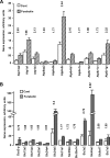
cAMP regulates the expression of vacuolar ATPases and ionic transporters. B16 mouse melanoma cells were incubated with or without (control) 20 μm forskolin for 36 h. RNA was purified, labeled, and analyzed on a pan-genomic mouse DNA microarray. A, ATPase expression was up-regulated after forskolin treatment by more than 70% (p < 0.05). B, SLC and P protein expression was stimulated after forskolin treatment by more than 70% (p < 0.05). -Fold change of stimulation is displayed above each histogram. Cont, control.
FIGURE 3.
FIGURE 3.
H89 prevents cAMP-induced melanosome alkalinization and melanin synthesis. B16 mouse melanoma cells were incubated with or without 20 μm forskolin (Fsk or control, respectively), in the presence or absence of 5 μm H89 for 36 h. A, cells were incubated with 30 μm DAMP for 30 min. After fixation in 3% paraformaldehyde and permeabilization with 0.1% Triton X-100, DAMP was visualized with fluorescein isothiocyanate-labeled anti-DNP, and melanosomes were labeled with anti-Tyrp1 (B8G3). Scale bar = 20 μm. B, representative cell pellets treated with vehicle (Cont), with forskolin, with H89 or with forskolin plus H89 (Fsk + H89) are displayed on the left panel. The melanin content, measured by spectrometry at 405 nm and normalized by the protein content, is shown on the right panel.
FIGURE 4.
FIGURE 4.
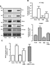
Effect of H89 on tyrosinase expression and activity. A, B16 mouse melanoma cells were incubated with or without 20 μm forskolin (Fsk or control, respectively), in presence or absence of 5 μm H89 for 36 h. Cells were solubilized, and proteins were analyzed by Western blot using antibodies against tyrosinase (Tyr), Tyrp1, Silver, Mitf, and Phospho-CREB (P-Creb) and, as loading control, Erk2. B, B16 mouse melanoma cells were transfected with a reporter plasmid, encoding luciferase under the control of either the tyrosinase promoter (P TYRO) or MITF promoter (P MITF). Cells were either mock-treated (control) or treated with 20 μm forskolin, in the presence or absence of 5 μm H89 for 36 h, and then solubilized and assayed for luciferase activity. C, B16 mouse melanoma cells were incubated with or without 20 μm forskolin (Fsk or control) in the presence or absence of 5 μm H89 for 36 h. Solubilized proteins were incubated with l-DOPA and assayed for DOPA oxidase activity by measuring the OD at 570 nm. To evaluate the direct effect of H89 on the DOPA oxidase activity of tyrosinase, H89 was added with l-DOPA from forskolin-treated cells (In vitro). Cont, control.
FIGURE 5.
FIGURE 5.
H89 affects melanosome structure. Shown are electron micrographs of B16 mouse melanoma cells incubated with or without 20 μm forskolin (Fsk or control, respectively) in the presence or absence of 5 μm H89 for 36 h. Scale bar = 1 μm. A higher magnification of melanosomes is shown in the lower left corner. Cont, control.
FIGURE 6.
FIGURE 6.
H89 blocks the melanogenic effects of LY294002 without inhibition of tyrosinase expression. B16 mouse melanoma cells were incubated with or without 20 μm forskolin or 5 μm LY294002 (Fsk, LY, or Cont, respectively), in the presence or absence of 5 μm H89 for 36 h. A, cells were solubilized, and proteins were analyzed by Western blotting using antibodies against tyrosinase (Tyr), Mitf and, as a loading control, ERK2. B, representative cell pellets are shown.
FIGURE 7.
FIGURE 7.
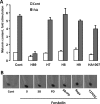
Neither PKA nor other kinases inhibited by H89 mediate the depigmenting effects of H89. A, B16 mouse melanoma cells were incubated with or without 20 μm forskolin (Fsk), in the presence of different PKA inhibitors (5 μm H89, 50 μm H7, 50 μm H8 and 50 μm H9, and 10 μm HA1007) for 36 h. Melanin content was measured by spectrometry at 405 nm and normalized by the protein concentration. B, B16 mouse melanoma cells were incubated with or without 20 μm forskolin, in the presence of different kinase inhibitors known to be inhibited by H89 for 36 h (SB202190 (SB), PD98059 (PD), Y27632, and rapamycin (Rapa)). Representative cell pellets are shown. Cont, control.

References

    1. Hearing V. J. (1999) J. Investig. Dermatol. Symp. Proc. 4, 24–28 - PubMed
    1. Hearing V. J. (2005) J. Dermatol. Sci. 37, 3–14 - PubMed
    1. Ortonne J. P. (2002) Br. J. Dermatol. 146, Suppl. 61, 7–10 - PubMed
    1. Buscà R., Ballotti R. (2000) Pigment Cell Res. 13, 60–69 - PubMed
    1. Cui R., Widlund H. R., Feige E., Lin J. Y., Wilensky D. L., Igras V. E., D'Orazio J., Fung C. Y., Schanbacher C. F., Granter S. R., Fisher D. E. (2007) Cell 128, 853–864 - PubMed

MeSH terms