. 2009 Jun 12;284(24):16562-16574.
doi: 10.1074/jbc.M109.010918. Epub 2009 Apr 22.

Identification and functional characterization of protein kinase A-catalyzed phosphorylation of potassium channel Kv1.2 at serine 449

Affiliations

Identification and functional characterization of protein kinase A-catalyzed phosphorylation of potassium channel Kv1.2 at serine 449

Rosalyn P Johnson et al. J Biol Chem. .

Abstract

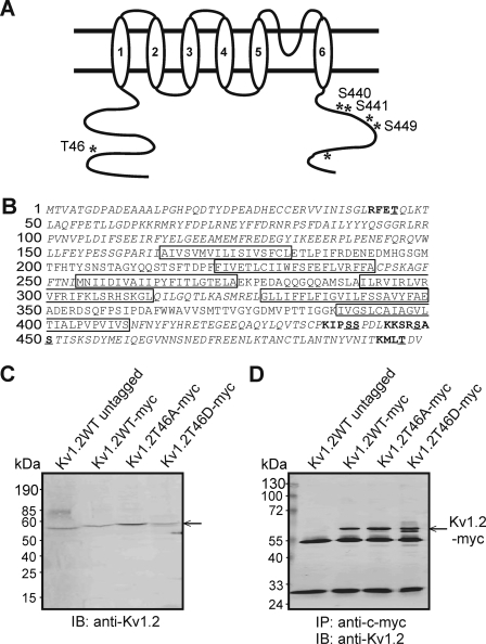
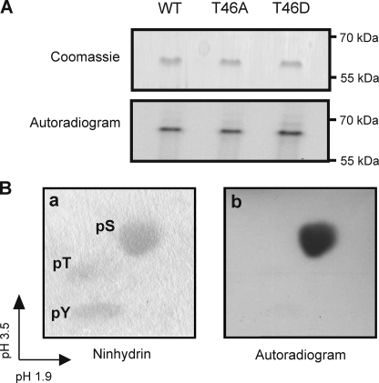
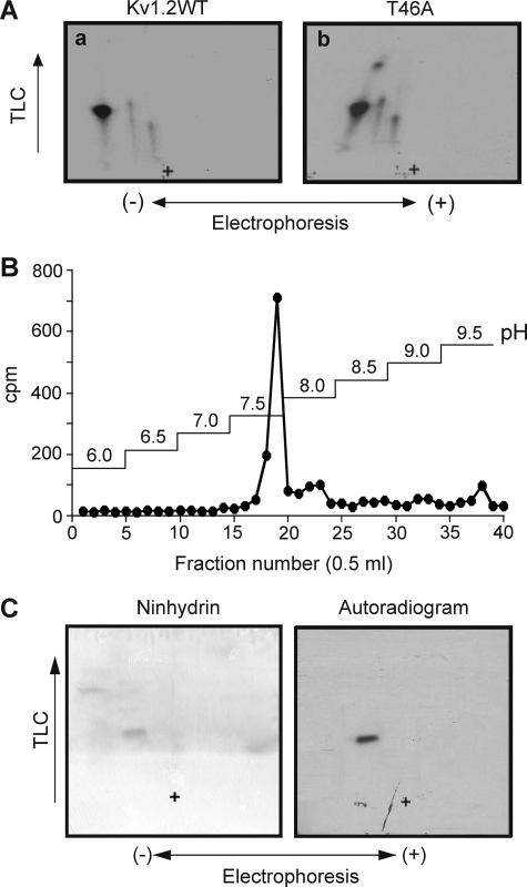
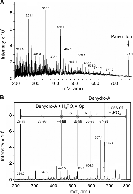
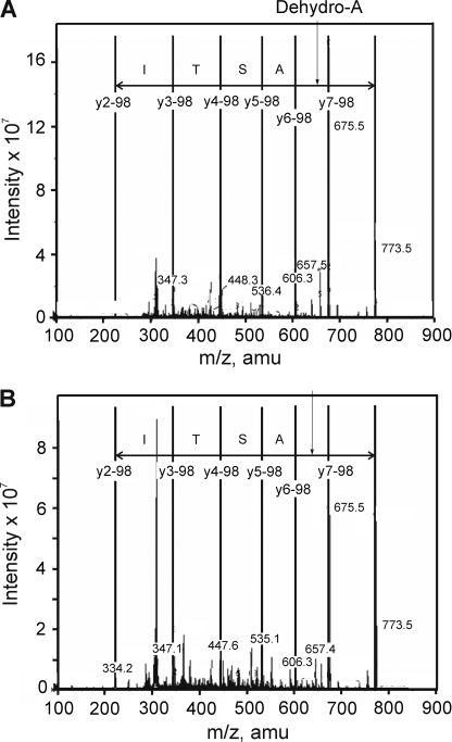
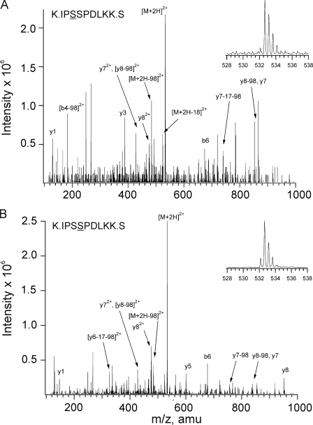
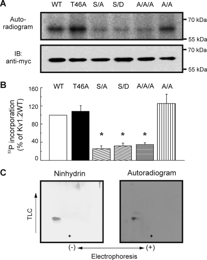
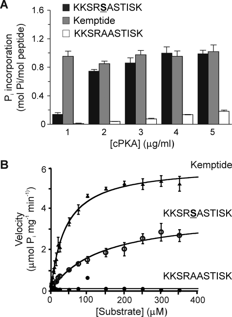
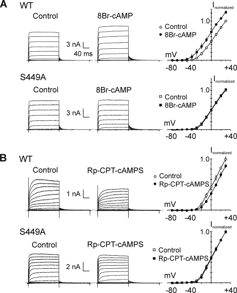
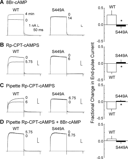
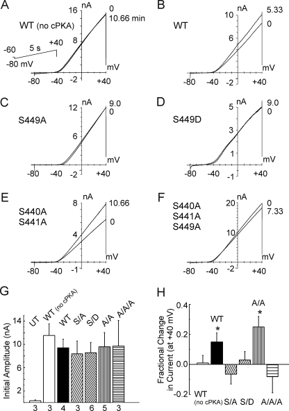
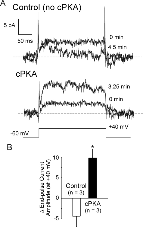
Vascular smooth muscle Kv1 delayed rectifier K+ channels (KDR) containing Kv1.2 control membrane potential and thereby regulate contractility. Vasodilatory agonists acting via protein kinase A (PKA) enhance vascule smooth muscle Kv1 activity, but the molecular basis of this regulation is uncertain. We characterized the role of a C-terminal phosphorylation site, Ser-449, in Kv1.2 expressed in HEK 293 cells by biochemical and electrophysiological methods. We found that 1) in vitro phosphorylation of Kv1.2 occurred exclusively at serine residues, 2) one major phosphopeptide that co-migrated with 449pSASTISK was generated by proteolysis of in vitro phosphorylated Kv1.2, 3) the peptide 445KKSRSASTISK exhibited stoichiometric phosphorylation by PKA in vitro, 4) matrix-assisted laser desorption ionization time-of-flight (MALDI-TOF) mass spectroscopy (MS) and MS/MS confirmed in vitro Ser-449 phosphorylation by PKA, 5) in situ phosphorylation at Ser-449 was detected in HEK 293 cells by MALDI-TOF MS followed by MS/MS. MIDAS (multiple reaction monitoring-initiated detection and sequencing) analysis revealed additional phosphorylated residues, Ser-440 and Ser-441, 6) in vitro 32P incorporation was significantly reduced in Kv1.2-S449A, Kv1.2-S449D, and Kv1.2-S440A/S441A/S449A mutant channels, but Kv1.2-S440A/S441A was identical to wild-type Kv1.2 (Kv1.2-WT), and 7) bath applied 8-Br-cAMP or dialysis with PKA catalytic subunit (cPKA) increased Kv1.2-WT but not Kv1.2-S449A current amplitude. cPKA increased Kv1.2-WT current in inside-out patches. Rp-CPT-cAMPS reduced Kv1.2-WT current, blocked the increase due to 8-Br-cAMP, but had no effect on Kv1.2-S449A. cPKA increased current due to double mutant Kv1.2-S440A/S441A but had no effect on Kv1.2-S449D or Kv1.2-S440A/S441A/S449A. We conclude that Ser-449 in Kv1.2 is a site of PKA phosphorylation and a potential molecular mechanism for Kv1-containing KDR channel modulation by agonists via PKA activation.


Figures

FIGURE 1.
FIGURE 1.
Identification of potential PKA consensus phosphorylation sites in Kv1.2 and immunoprecipitation of Myc-tagged wild-type and mutant Kv1.2. A, schematic of Kv1.2 with candidate PKA phosphorylation sites indicated by asterisks, and Ser/Thr residues relevant to the study are labeled. B, amino acid sequence of Kv1.2 indicating transmembrane domains (boxes), cytoplasmic domains (italics) assessed by MIDAS for phosphorylated residues, candidate PKA consensus sites (bold), and potential phosphorylated Ser/Thr residues (underlined). C, immunoblot (IB) analysis of untagged Kv1.2-WT and Myc-tagged WT and mutant constructs using anti-Kv1.2. D, immunoblot identification of untagged Kv1.2-WT and Myc-tagged WT and mutant constructs by anti-Kv1.2 in anti-Myc immunoprecipitates (IP).
FIGURE 2.
FIGURE 2.
In vitro phosphorylation of Kv1.2 serine residue(s) by PKA. A, Kv1.2-WT-myc, -T46A-myc, and -T46D-myc immunoprecipitates treated with cPKA in the presence of [γ-32P]ATP and visualized by Coomassie Blue staining and autoradiography to detect 32P incorporation. Data are representative of six experiments. B, phosphoamino acid analysis of phosphorylated Kv1.2-WT-myc protein by two-dimensional electrophoresis. Unlabeled phosphoserine (pS), phosphothreonine (pT), and phosphotyrosine (pY) standards were detected by ninhydrin staining (a) and 32P-labeled residues by autoradiography (b). Data are representative of four experiments.
FIGURE 3.
FIGURE 3.
Phosphopeptide mapping and affinity purification of Kv1.2-WT and T46A phosphopeptides and pSASTISK co-migrates with tryptic phosphopeptide derived from PKA-phosphorylated Kv1.2-WT. A, phosphopeptide maps of Kv1.2-WT-myc (a) and -T46A-myc (b) phosphorylated by cPKA in the presence of [γ-32P]ATP. Data are representative of six independent experiments. + indicates the origin. B, radioactivity profile of PKA-phosphorylated Kv1.2-WT-myc tryptic peptides separated by immobilized metal affinity chromatography. C, pSASTISK was mixed with the tryptic digest of 32P-phosphorylated Kv1.2-WT-myc and separated by electrophoresis and chromatography. Synthetic phosphopeptide (left) and 32P-labeled phosphopeptides (right) were visualized by ninhydrin staining and autoradiography, respectively. + indicates the origin.
FIGURE 4.
FIGURE 4.
Identification of Ser-449 of Kv1.2 as a site of phosphorylation after in vitro PKA treatment. 32P-Labeled tryptic peptides of Kv1.2-WT-myc after in vitro PKA treatment were partially purified on a C18 column and analyzed by mass spectrometry. A, ion trap full scan MS spectrum of Kv1.2-myc tryptic digest showing a major phosphopeptide with m/z 773.4. B, MS/MS spectrum resulting from isolation and collision-induced fragmentation of the singly charged m/z 773.4 precursor ion. The sequence of the m/z 773.4 ion shown is based on a y-ion series (separated by vertical lines). amu, atomic mass units; Sp, phosphoserine.
FIGURE 5.
FIGURE 5.
Confirmation of in situ phosphorylation of Kv1.2 at Ser-449. A, MS/MS spectrum resulting from isolation and collision-induced fragmentation of the singly charged m/z 773.4 precursor ion obtained from ion trap full scan MS spectrum of control HEK 293 cell Kv1.2-WT-myc tryptic digest. The sequence of the m/z 773.4 ion shown is based on a y-ion series (separated by vertical lines). B, MS/MS spectrum of the singly charged m/z 773.4 precursor ion obtained from ion trap full scan MS spectrum of 8-Br-cAMP-treated HEK 293 cell Kv1.2-WT-myc tryptic digest. The sequence is as indicated in A. amu, atomic mass units.
FIGURE 6.
FIGURE 6.
Identification of Ser-440 and Ser-441 as additional sites of in situ phosphorylation of Kv1.2. MS/MS spectra obtained from MIDAS analysis of tryptic digest of control HEK 293 cell Kv1.2 with ion assignments based on Mascot annotation and manual validation. A, spectrum of early eluting phosphopeptide. B, spectrum of late eluting phosphopeptide. Insets in each panel represent enhanced resolution scans of the respective precursor ions (m/z 532.7). Identical spectra were obtained for 8-Br-cAMP-treated cells expressing Kv1.2-WT. amu, atomic mass units.
FIGURE 7.
FIGURE 7.
Quantitative analysis of 32P incorporation by wild-type and mutant Kv1.2 constructs during in vitro PKA treatment. A, levels of 32P incorporation and protein in anti-Myc immunoprecipitates of tagged Kv1.2-WT (WT), -T46A, -S449A (S/A), -S449D (S/D), -S440A/S441A (A/A), and -S440A/S441A/S449A (A/A/A) channels after in vitro phosphorylation by PKA were detected by autoradiography (upper panel) and immunoblotting (IB) with anti-Myc (lower panel), respectively. B, 32P incorporation relative to total protein was determined by densitometric analysis and expressed as a % of the value for Kv1.2-WT. Data are the means ± S.E. (n = 5). Asterisks indicate significant difference (p < 0.05) from Kv1.2-WT by analysis of variance followed by the Bonferroni post-hoc test. C, co-migration of synthetic pSASTISK and 32P-labeled Kv1.2-S449A-myc mutant tryptic phosphopeptides was detected by ninhydrin staining and autoradiography. Data are representative of three experiments. + indicates the origin.
FIGURE 8.
FIGURE 8.
Stoichiometric and kinetic analysis of PKA-catalyzed 32P incorporation by synthetic peptide. Synthetic peptide KKSRSASTISK, corresponding to amino acid residues 445–455 of Kv1.2 and containing the putative PKA site Ser-449, was compared as a substrate to LLRRASLG (Kemptide) (positive control) and KKSRAASTISK (corresponding to the sequence of Kv1.2-S449A mutant, negative control). A, fixed concentrations of each peptide (50 μm) were treated with [γ-32P]ATP and cPKA (1.0–5.0 μg/ml), and 32P incorporation was quantified by C̆erenkov counting. Data are the means ± S.E. of three independent experiments carried out in triplicate. B, cPKA activity was determined by quantification of 32P incorporation into KKSRSASTISK, Kemptide, and KKSRAASTISK peptides during the linear phase of the reaction (t = 1.5 min). Enzyme velocities were plotted against substrate concentration and fitted to the Michaelis-Menten equation to obtain Km and Vmax values.
FIGURE 9.
FIGURE 9.
S449A mutation prevents modulation of Kv1.2 current amplitude by PKA activation or inhibition. A, representative families of whole-cell currents recorded during 200-ms pulses between −80 and +30 mV in 10-mV steps from HEK 293 cells transiently expressing Kv1.2-WT (WT; n = 4) or Kv1.2-S449A (S449A; n = 4) before (Control) and after 8-Br-cAMP (1 mm; 8-Br-cAMP) treatment and the corresponding mean (±S.E.) fractional I-V relations in control (open circles) and 8-Br-cAMP (closed circles) (i.e. plots are the average values of end pulse current at each voltage ± 8-Br-cAMP normalized to value in control condition at +30 mV in each cell). Mean fractional current in 8-Br-cAMP was significantly greater than that in control condition at all voltages positive to −40 mV for Kv1.2-WT (p < 0.05; unpaired Student's t test) but not different for Kv1.2-S449A. B, representative families of Kv1.2-WT (WT; n = 4) or Kv1.2-S449A (S449A; n = 3) whole-cell currents and corresponding mean ± S.E. fractional I-V relations (as in A) before (Control) and after Rp-CPT-cAMPS (25 μm; Rp-CPT-cAMPS) treatment. Mean fractional current in Rp-CPT-cAMPS was significantly reduced compared with control conditions at all voltages positive to −20 mV for Kv1.2-WT (p < 0.05; unpaired Student's t test) but not different for Kv1.2-S449A.
FIGURE 10.
FIGURE 10.
Time dependence of Kv1.2 current amplitude modulation by PKA activation and inhibition and effect of Rp-CPT-cAMPS on basal and 8-Br-cAMP-induced increase in Kv1.2 current. A, representative whole-cell currents during 250-ms voltage steps to +20 mV (applied at 15-s intervals) recorded before (0 min) and when a stable change in current amplitude was detected at 4 min for Kv1.2-WT (WT; n = 3) and at 14 min for Kv1.2-S449A (S449A; n = 4) as well as the mean (±S.E.) fractional change in Kv1.2-WT (n = 3) and S449A (n = 4) end pulse current (n = 4) due to treatment with 8-Br-cAMP (1 mm). B, representative whole-cell currents (evoked by voltage protocol in A) recorded before (0 min) and when a stable change in current amplitude was detected at 5.75 min for Kv1.2-WT (WT) and at 6 min for Kv1.2-S449A (S449A) as well as the mean (±S.E.) fractional change in Kv1.2-WT (n = 4) and S449A (n = 3) end pulse current due to bath treatment with Rp-CPT-cAMPS (25 μm). C, representative Kv1.2-WT and Kv1.2-S449A whole-cell currents (evoked by protocol in A) recorded immediately after membrane rupture (0 min) and after 6 and 6.75 min, respectively, of dialysis with Rp-CPT-cAMPS (25 μm) as well as the mean (±S.E.) fractional change in Kv1.2-WT (n = 4) and S449A (n = 3) end pulse current due to Rp-CPT-cAMPS. D, representative Kv1.2-WT and Kv1.2-S449A whole-cell currents (evoked by protocol in A) before (0) and after 5.75 and 6.75 min of treatment with 8-Br-cAMP (1 mm), respectively, after dialysis with Rp-CPT-cAMPS (25 μm) as well as the mean (±S.E.) fractional change in end pulse current (n = 3 in each group) due to 8-Br-cAMP in the presence of Rp-CPT-cAMPS. The asterisks in A–D indicate values for S449A mutant were significantly different from Kv1.2-WT by unpaired Student's t test (p < 0.05).
FIGURE 11.
FIGURE 11.
Modulation of whole-cell Kv1.2 current by cPKA requires Ser-449. A–F, representative whole-cell currents due to wild-type (WT) and mutant Kv1.2 (S449A, S449D, S440A/S441A, and S440A/S441A/S449A) constructs evoked by 5-s voltage ramps from −80 to +40 mV (applied at 20-s intervals) immediately after membrane rupture (0 min) and after the indicated times of dialysis with control pipette solution (panel A; Kv1.2WT (WT (no cPKA)) or solution with cPKA (50 nm; B–F). The second tracing in each panel represents the stable level of current achieved after the indicated time. G, current density at +40 mV immediately after membrane rupture ±S.E. for untransfected HEK 293 cells (UT) and cells expressing Kv1.2-WT (WT (no cPKA) and WT), -S449A (S/A), -S449D (S/D), -S440A/S441A (A/A), or -S440A/S441A/S449A (A/A/A) mutant constructs used in A–F (numbers indicate n cells from >3 transfections). H, mean fractional change in current at +40 mV ± S.E. (evoked by the ramp protocol indicated in A) for cells expressing wild-type or mutant Kv1.2 constructs after dialysis with control pipette solution (WT (no cPKA)) or solution containing cPKA (Kv1.2-WT (WT), S449A (S/A), S449D (S/D), S440A/S441A (A/A), and S440A/S441A/S449A (A/A/A)) (n values are as in G). The asterisks indicate significantly different (p < 0.05) from Kv1.2-WT in the absence of PKA (i.e. WT (no cPKA)) determined by analysis of variance followed by the Bonferroni post-hoc test.
FIGURE 12.
FIGURE 12.
Modulation of Kv1.2-WT current in excised, inside-out membrane patches by cPKA. A, representative recordings of membrane patch current before (0 min) and after 4.5 and 3.25 min in control conditions and solutions containing 50 nm cPKA, respectively. B, average stable change in end-pulse current amplitude in control bath solution and in the presence of cPKA after 3.5–4.5 min (n = 3 patches each). The asterisk indicates significantly different from control, determined by unpaired Student's t test (p < 0.05).

Similar articles

Cited by

References

    1. Nelson M. T., Quayle J. M. ( 1995) Am. J. Physiol. 268, C799– C822 - PubMed
    1. Knot H. J., Nelson M. T. ( 1995) Am. J. Physiol. 269, H348– H355 - PubMed
    1. Davis M. J., Hill M. A. ( 1999) Physiol. Rev. 79, 387– 423 - PubMed
    1. Kerr P. M., Clément-Chomienne O., Thorneloe K. S., Chen T. T., Ishii K., Sontag D. P., Walsh M. P., Cole W. C. ( 2001) Circ. Res. 89, 1038– 1044 - PubMed
    1. Thorneloe K. S., Chen T. T., Kerr P. M., Grier E. F., Horowitz B., Cole W. C., Walsh M. P. ( 2001) Circ. Res. 89, 1030– 1037 - PubMed

Publication types