. 2009 Jul;297(1):G135-43.
doi: 10.1152/ajpgi.90386.2008. Epub 2009 Apr 23.

HCl-activated neural and epithelial vanilloid receptors (TRPV1) in cat esophageal mucosa

Affiliations

HCl-activated neural and epithelial vanilloid receptors (TRPV1) in cat esophageal mucosa

Ling Cheng et al. Am J Physiol Gastrointest Liver Physiol. 2009 Jul.

Abstract

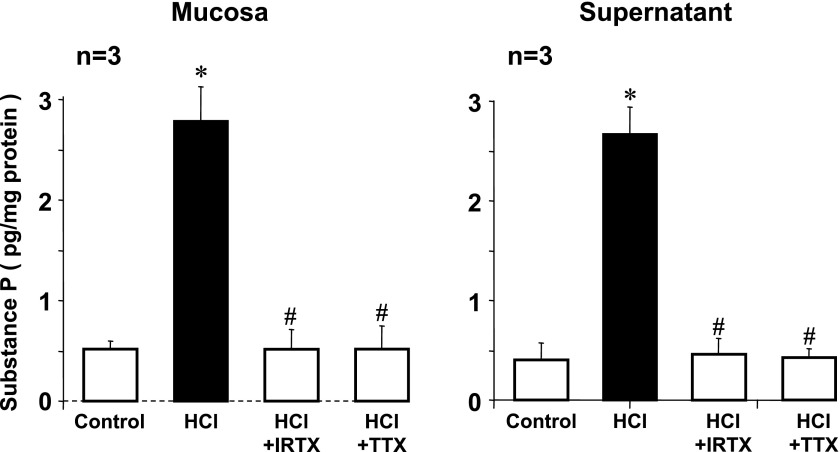
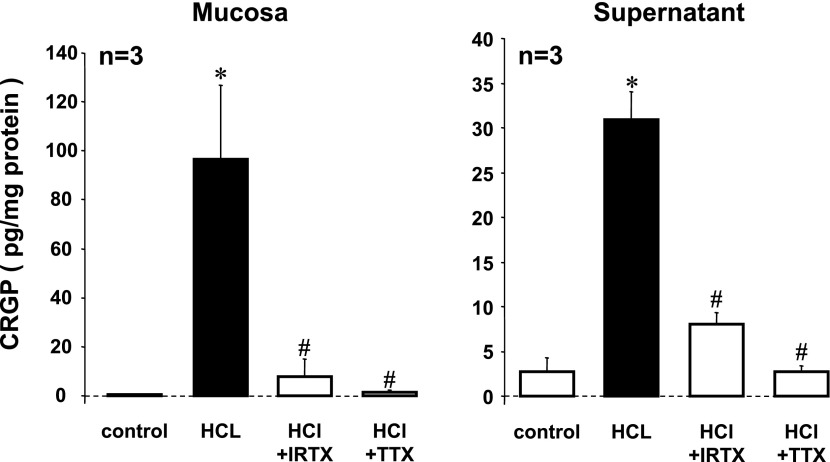
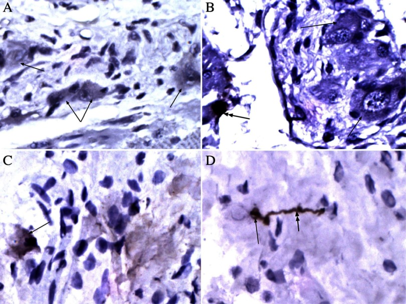
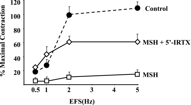
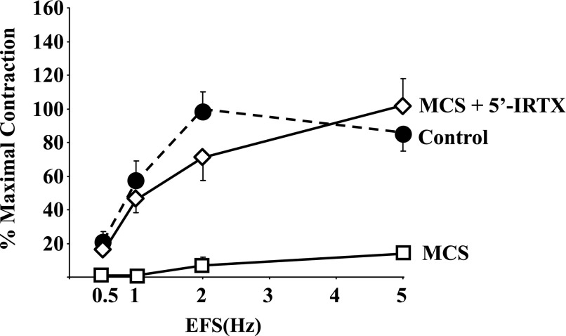
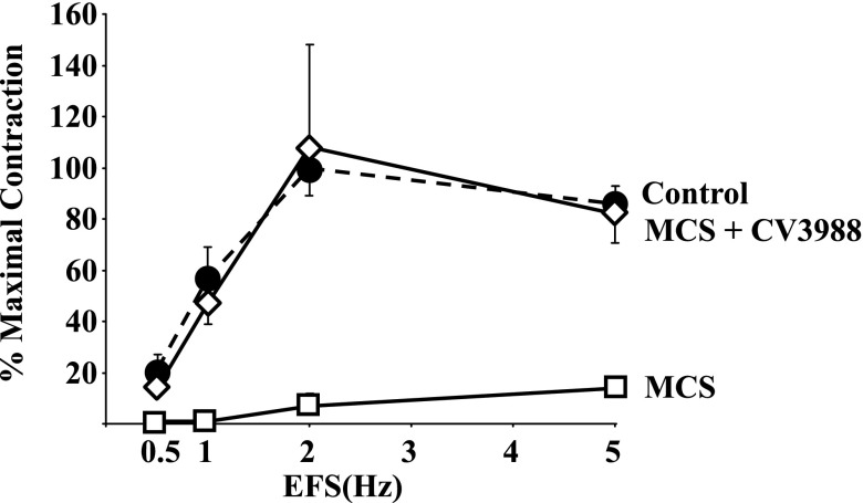
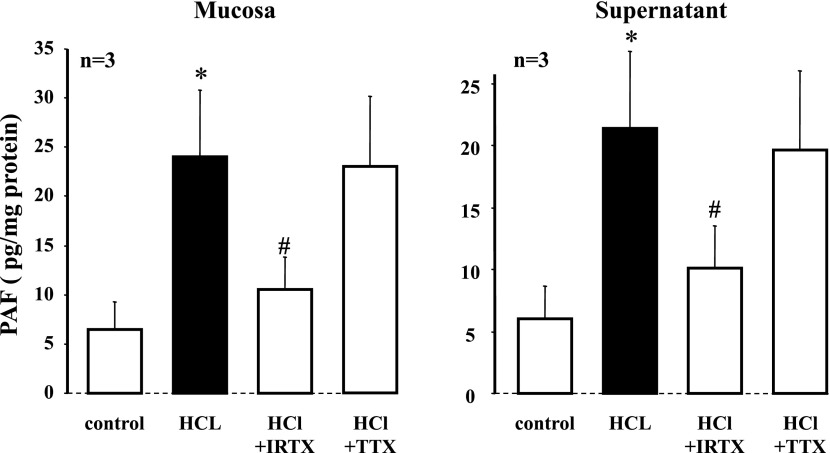
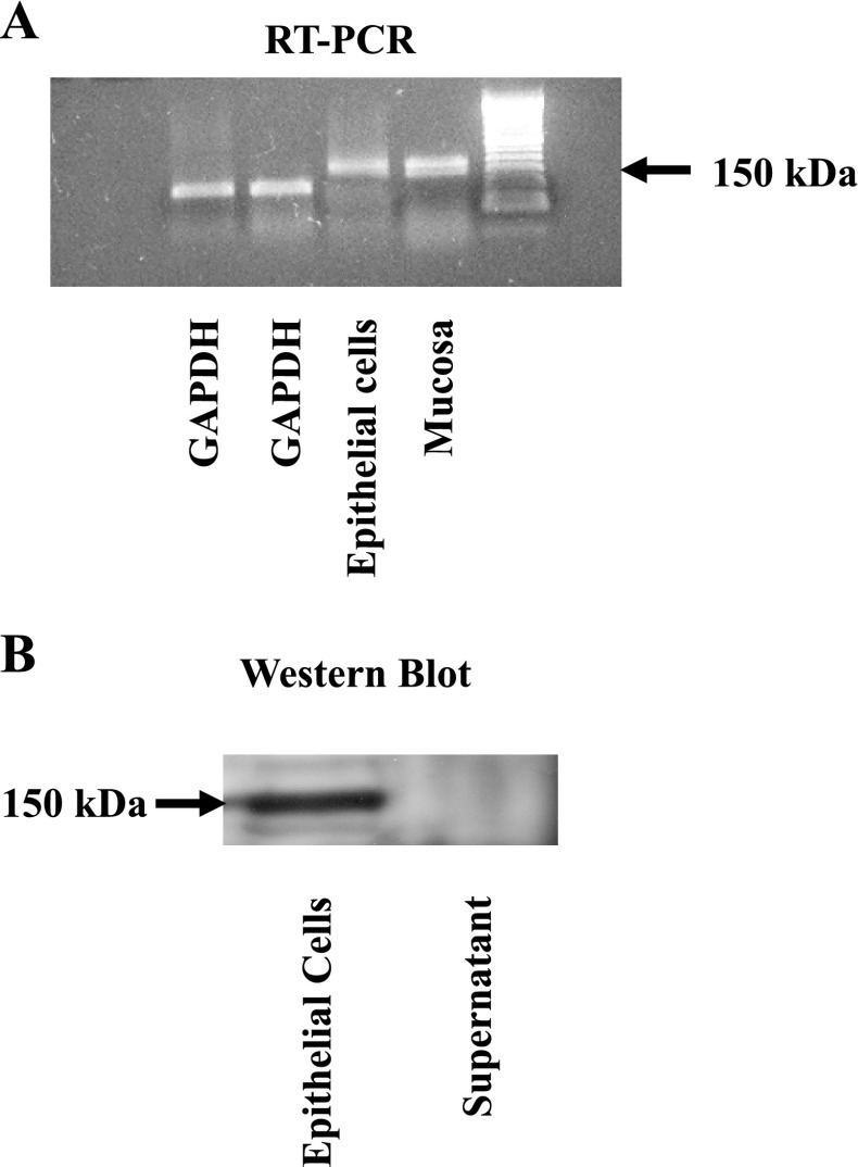
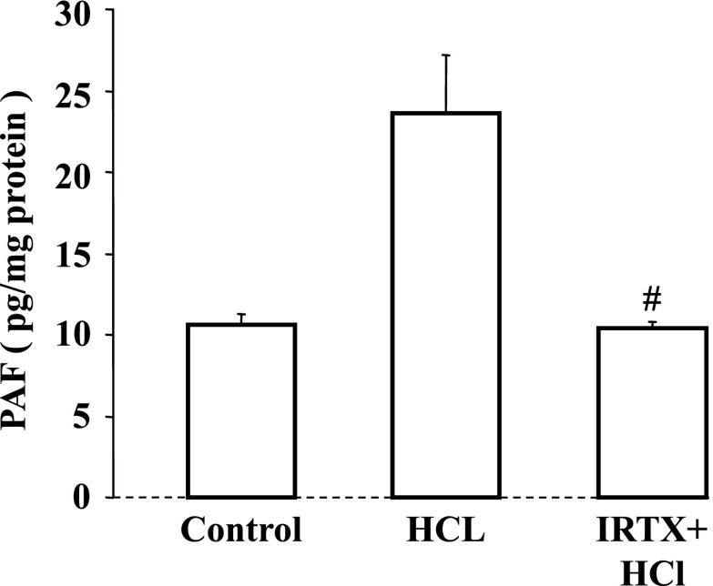
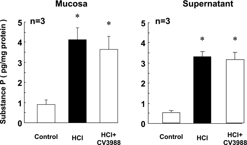
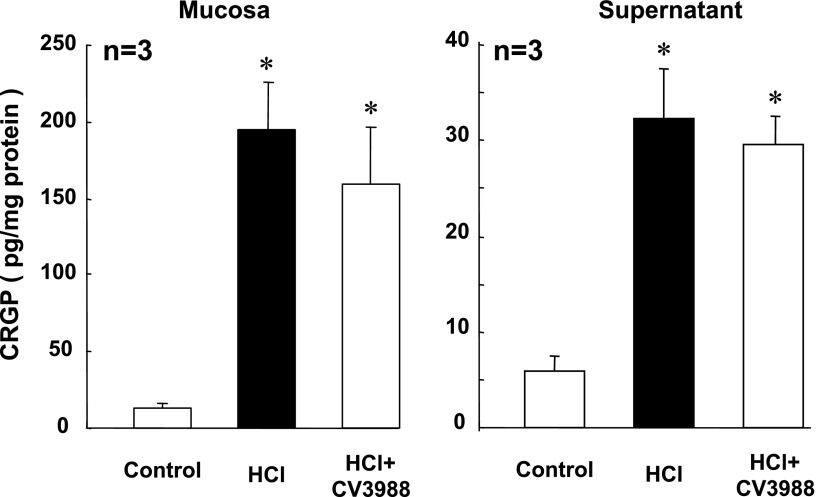
To test whether transient receptor potential channel vanilloid subfamily member-1 (TRPV1) mediates acid-induced inflammation in the esophagus, a tubular segment of esophageal mucosa was tied at both ends, forming a sac. The sac was filled with 0.01 N HCl (or Krebs buffer for control) and kept in oxygenated Krebs buffer at 37 degrees C. The medium around the sac (supernatant) was collected after 3 h. Supernatant of the HCl-filled sac abolished contraction of esophageal circular muscle strips in response to electric field stimulation. Contraction was similarly abolished by supernatant of mucosal sac filled with the TRPV1 agonist capsaicin (10(-6) M). These effects were reversed by the selective TRPV1 antagonist 5'-iodoresiniferatoxin (IRTX) and by the platelet-activating factor (PAF) receptor antagonist CV9388. Substance P and CGRP levels in mucosa and in supernatant increased in response to HCl, and these increases were abolished by IRTX and by tetrodotoxin (TTX) but not affected by CV9388, indicating that substance P and CGRP are neurally released and PAF independent. In contrast, the increase in PAF was blocked by IRTX but not by TTX. Presence of TRPV1 receptor was confirmed by RT-PCR and by Western blot analysis in whole mucosa and in esophageal epithelial cells enzymatically isolated and sorted by flow cytometry or immunoprecipitated with cytokeratin antibodies. In epithelial cells PAF increased in response to HCl, and the increase was abolished by IRTX. We conclude that HCl-induced activation of TRPV1 receptors in esophageal mucosa causes release of substance P and CGRP from neurons and release of PAF from epithelial cells.


Figures

Fig. 1.
Fig. 1.
When in vitro mucosa is exposed to HCl for 3 h, substance P levels increase 6 times in the mucosa and in the supernatant compared with control (pH 7.4) (*P < 0.05 ANOVA). The HCl-induced increase in substance P is abolished by the transient receptor potential channel vanilloid-1 (TRPV1) receptor antagonist 5′-iodoresiniferatoxin (IRTX) and by the neural blocker tetrodotoxin (TTX) both in the mucosa and in the supernatant (#P < 0.05 ANOVA). Bars represent means ± SE of mucosal tissue from 3 animals.
Fig. 2.
Fig. 2.
When in vitro mucosa is exposed to HCl for 3 h, calcitonin gene-related peptide (CGRP) levels increase 200 times in the mucosa and 10 times in the supernatant compared with control (pH 7.4) (*P < 0.05 ANOVA). The HCl-induced increase in CGRP is abolished by the TRPV1 receptor antagonist IRTX and by the neural blocker TTX both in the mucosa and in the supernatant (#P < 0.05 ANOVA). Bars represent means ± SE of mucosal tissue from 3 animals.
Fig. 3.
Fig. 3.
Histological sections of esophagus immunostained to detect CGRP (A, B) or substance P (C, D). Immunoreactivity was detected with secondary antibody tagged with horseradish peroxidase-conjugated polymer and diaminobenzidine (brown precipitate). Sections were counterstained with hematoxylin (blue) to depict unlabeled cells. A and B: CGRP immunoreactivity was localized in ganglion cells (note nucleoli). In general, in the submucous plexus the immunoreactivity was low level and distributed throughout the cytoplasm (arrows), although occasional cells had intense levels of CGRP immunoreactivity (double-headed arrow, B). C and D: substance P immunoreactivity was easily detected in scattered small neurons and fibers of the submucous plexus. In addition, fine substance P-immunoreactive fibrils were distributed in the submucosa, but occasional longer and somewhat tortuous fibers (probably axons) were identified (double-headed arrow, D).
Fig. 4.
Fig. 4.
Contraction of esophageal circular muscle (Control) in response to electrical field stimulation (EFS) is almost abolished by supernatant of HCl-filled mucosal sac (MHS). The reduction is reversed by application of the TRPV1 antagonist IRTX (10−5 M) inside the mucosal sac (MHS+5′-IRTX), confirming presence of TRPV1 vanilloid receptors in the mucosal wall. Data represent means ± SE of muscle strips from 3 animals.
Fig. 5.
Fig. 5.
Contraction of esophageal circular muscle (Control) in response to EFS is almost abolished by supernatant of capsaicin-filled mucosal sac (MCS). The reduction is reversed by application of the TRPV1 antagonist IRTX (10−5 M) inside the mucosal sac (MCS+5′-IRTX), confirming presence of TRPV1 vanilloid receptors in the mucosal wall. Data represent means ± SE of muscle strips from 3 animals.
Fig. 6.
Fig. 6.
EFS-induced contraction of esophageal circular muscle (Control) is almost abolished by supernatant of capsaicin (10−6 M)-filled mucosal sac (MCS). The reduction is reversed by preexposure of the muscle to the platelet-activating factor (PAF) receptor antagonist CV3988 (MCS+CV3988; 10−6 M). This is consistent with release of PAF by mucosa in response to activation of TRPV1 by capsaicin and suggests that PAF may be the inflammatory mediator most responsible for impairment of EFS-induced (i.e., neurally induced) contraction of circular muscle. Data represent means ± SE of muscle strips from 3 animals.
Fig. 7.
Fig. 7.
When in vitro mucosa is exposed to HCl for 3 h, PAF levels increase 4–5 times in the mucosa and in the supernatant compared with control (pH 7.4) (*P < 0.05 ANOVA). The HCl-induced increase in PAF is abolished by the TRPV1 receptor antagonist IRTX (#P < 0.05 ANOVA) and not affected by the neural blocker TTX both in the mucosa and in the supernatant, indicating that production of PAF does not involve axonal conductance. Data represent means ± SE of mucosal tissue from 3 animals.
Fig. 8.
Fig. 8.
A: to demonstrate the presence of TRPV1 receptor mRNA in nonneural cells, esophageal epithelial cells were enzymatically isolated and sorted by flow cytometry. Primers were derived from conserved regions of mRNA sequences of human, rat, dog, mouse, guinea pig, and rabbit. RT-PCR confirmed the presence of TRPV1 receptor mRNA in these cat esophageal epithelial cells. B: to perform Western blot analysis, epithelial cells were immunoprecipitated with an epithelial cell-selective pan-cytokeratin antibody conjugated with magnetic beads and were isolated by exposing the suspension to a magnetic field as specified by the manufacturer. A 150-kDa band was immunoblotted with a TRPV1 antibody, confirming the presence of TRPV1 receptors in these cat esophageal epithelial cells.
Fig. 9.
Fig. 9.
In enzymatically isolated epithelial cells sorted to 95% purity by flow cytometry, 3-h exposure to HCl more than doubled PAF levels (#P < 0.05 ANOVA) The increase in PAF was abolished by the TRPV1 receptor antagonist IRTX (10−5 M). Data represent means ± SE of epithelial cells from 3 animals.
Fig. 10.
Fig. 10.
When in vitro mucosa is exposed to HCl for 3 h, substance P levels significantly increase in the mucosa and in the supernatant compared with control (pH 7.4) (*P < 0.05 ANOVA). The HCl-induced increase in substance P was not affected by the PAF receptor antagonist CV3988. Bars represent means ± SE of mucosal tissue from 3 animals.
Fig. 11.
Fig. 11.
When in vitro mucosa is exposed to HCl for 3 h, CGRP levels significantly increase in the mucosa and in the supernatant compared with control (pH 7.4) (*P < 0.05 ANOVA). The HCl-induced increase in CGRP was not affected by the PAF receptor antagonist CV3988. Bars represent means ± SE of mucosal tissue from 3 animals.

Similar articles

Cited by

References

    1. Barbara G, De Giorgio R, Stanghellini V, Corinaldesi R, Cremon C, Gerard N, Gerard C, Grady EF, Bunnett NW, Blennerhassett PA, Collins SM. Neutral endopeptidase (EC 3.42411) downregulates the onset of intestinal inflammation in the nematode infected mouse. Gut 52: 1457–1464, 2003. - PMC - PubMed
    1. Bartsch I, Zschaler I, Haseloff M, Steinberg P. Establishment of a long-term culture system for rat colon epithelial cells. In Vitro Cell Dev Biol Anim 40: 278–284, 2004. - PubMed
    1. Behar J, Guenard V, Walsh JH, Biancani P. VIP and acetylcholine: neurotransmitters in esophageal circular smooth muscle. Am J Physiol Gastrointest Liver Physiol 257: G380–G385, 1989. - PubMed
    1. Bhalla V, Liu J, Mittal R. Nature of symptoms induced by vanilloid receptors activation in the human esophagus (Abstract). Gastroenterology 126: A111, 2004.
    1. Bhat YM, Bielefeldt K. Capsaicin receptor (TRPV1) and non-erosive reflux disease. Eur J Gastroenterol Hepatol 18: 263–270, 2006. - PubMed

Publication types

MeSH terms