. 2009 Jun;174(6):2107-15.
doi: 10.2353/ajpath.2009.081018. Epub 2009 Apr 23.

Requirement of the Akt/beta-catenin pathway for uterine carcinosarcoma genesis, modulating E-cadherin expression through the transactivation of slug

Affiliations

Requirement of the Akt/beta-catenin pathway for uterine carcinosarcoma genesis, modulating E-cadherin expression through the transactivation of slug

Makoto Saegusa et al. Am J Pathol. 2009 Jun.

Abstract

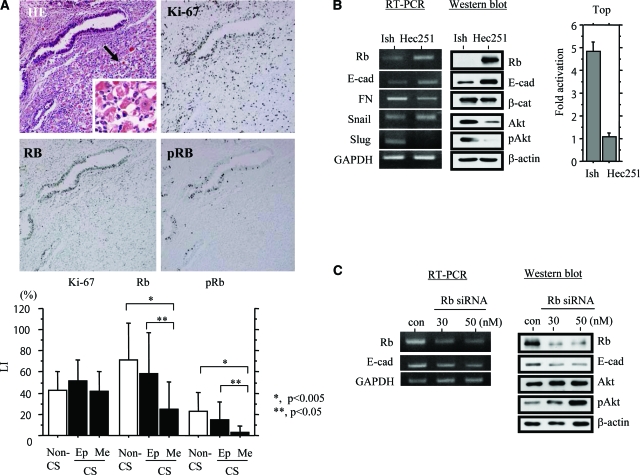
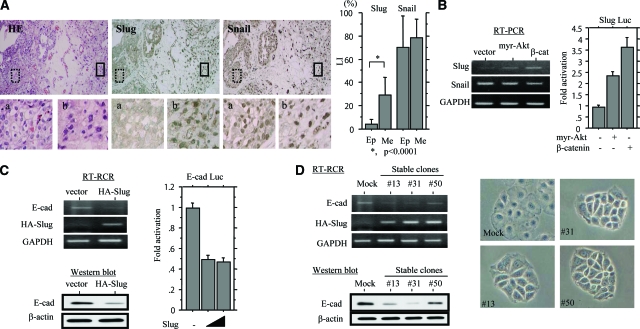
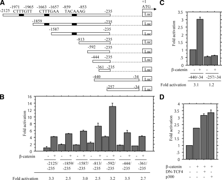
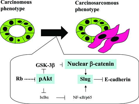
Uterine carcinosarcomas (UCSs) are considered to represent true examples of the epithelial-mesenchymal transition. Akt plays a key role in the induction of epithelial-mesenchymal transition, but little is known about its involvement in tumorigenesis. Here we examined the functional roles of the Akt/beta-catenin pathway in UCSs. In clinical samples, phospho-Akt (pAkt) expression was found to be significantly increased in mesenchymal compared with epithelial components, exhibiting both positive and negative correlations with nuclear beta-catenin and E-cadherin, respectively. Expression levels of the transcription factor Slug were also significantly up-regulated in the mesenchymal components and strongly correlated with both pAkt and nuclear beta-catenin. In endometrial cancer cell lines, active Akt induced the stabilization of nuclear beta-catenin through the phosphorylation of GSK-3beta, and this, in turn, led to the transactivation of Slug, which was mediated by nuclear beta-catenin. Moreover, Slug overexpression itself caused repression of E-cadherin, with subtle changes in cell morphology. In addition, knockdown of the retinoblastoma gene product (Rb) up-regulated pAkt and repressed E-cadherin, consistent with the in vivo finding of significantly decreased Rb expression in mesenchymal components. These findings suggest that changes in the Akt/beta-catenin pathway, as well as alterations in Rb expression, may be essential for both the establishment and maintenance of phenotypic characteristics of UCSs, playing key roles in the regulation of E-cadherin through the transactivation of the Slug gene.


Figures

Figure 1
Figure 1
Expression of Rb and pRb in UCSs. A, Top: H&E staining and immunohistochemistry for Rb, pRb, and Ki-67 are illustrated. The mesenchymal lesion indicated by the arrow is magnified in the inset. Bottom: Nuclear LIs for Rb, pRb, and Ki-67 in epithelial (Ep) and mesenchymal (Me) components of uterine CS (carcinosarcoma), as well as non-CS tumors. B, Left and middle: Comparison of expression levels of several EMT-related molecules for mRNA by RT-PCR (left) and protein by Western blot (middle) between Ishikawa (Ish) and Hec251 cells. E-cad, E-cadherin; FN, fibronectin Right: Ishikawa and Hec251 cells were transfected with Top reporter constructs to determine β-catenin-dependent transcriptional activity. Relative activity was determined based on arbitrary light units of luciferase activity normalized to pRL-TK activity. The activities of the reporter plus the effector relative to that of the reporter plus empty vector are shown as means ± SD. C: Analysis of mRNA (at 24 hours after transfection) (left) and total protein (at 48 hours) (right) in Hec251 cells transfected 30 and 50 nmol/L siRNAs for Rb. A siRNA with no homology to the mammalian genome was used as a negative control (con). Original magnifications: ×100 (A, top); ×400 (A, inset).
Figure 2
Figure 2
Akt regulates expression of E-cadherin and β-catenin. A: Serial sections through a UCS. H&E and immunohistochemistry for E-cadherin, pAkt, and β-catenin are illustrated. The lesions enclosed by boxes in the top panels are magnified in the bottom panels. B: Immunohistochemical (IHC) scores for E-cadherin (left) and LIs for pAkt and nuclear (N) β-catenin (right) in epithelial (Ep) and mesenchymal (Me) components of uterine CS (carcinosarcoma), as well as non-CS tumors. C, Top left: Western blot analysis of E-cadherin (E-cad) was performed with subcellular protein fractions (Cyt, cytoplasmic; Nu, nuclear) extracted from myr-Akt-transfected Hec251 cells. In the pAkt panel, upper bands (indicated by the asterisk) are exogenous myr-Akt, in contrast to the lower bands demonstrating endogenous pAkt. Bottom left: After transfection of myr-Akt into Hec251 cells, staining for pAkt and E-cadherin was performed. Note the lack of E-cadherin expression in cell overexpressing myr-Akt (indicated by arrows). Top right: Analysis of E-cadherin (E-cad) mRNA levels by RT-PCR with total RNA extracted from myr-Akt-transfected Hec251 cells. Bottom right: Hec251 cells were transfected with reporter constructs containing E-cadherin promoter (E-cad Luc), together with expression plasmid for myr-Akt. Relative activity was determined based on arbitrary light units of luciferase activity normalized to pRL-TK activity. The activities of the reporter plus the effector relative to that of the reporter plus empty vector are shown as means ± SD. D, Top left: Western blot analysis of HA-β-catenin, total GSK-3β, and phospho-GSK-3β (pGSK-3β) was performed with subcellular protein fractions (Cyt, cytoplasmic; Nu, nuclear) extracted from myr-Akt-transfected Ish-bcat mo.#25 cells. Bottom left: After transfection of myr-Akt, Ish-bcat#25 cells were treated with cycloheximide (25 μg/ml) for the times shown. Western blotting was performed using anti-HA, anti-pAkt, anti-GSK-3β, and anti-pGSK-3β antibodies. In the pAkt panel, upper bands (indicated by the asterisk) are exogenous myr-Akt, in contrast to lower bands demonstrating endogenous pAkt. Top and bottom right: Relative amounts of HA-β-catenin were calculated by normalization to the signals for β-actin, using NIH Image. Expression levels of HA-β-catenin observed in myr-Akt-transfected cells in the absence of cycloheximide treatment (0 hours) were set as 100%. The experiment was performed in triplicate. Original magnifications: ×100 (A, top); ×400 (A, bottom).
Figure 3
Figure 3
Up-regulation of Slug in UCSs. A, Left: Serial sections through a UCS. H&E staining and immunohistochemistry for Slug and Snail are illustrated. Boxes with dotted and solid outlines enclose epithelial and mesenchymal lesions magnified in a and b, respectively. Right: Nuclear LIs for Slug and Snail in epithelial (Ep) and mesenchymal (Me) components of UCSs. B, Left: Analysis of mRNA levels for Slug and Snail by RT-PCR with total RNA extracted from Hec251 cells transfected with myr-Akt and β-catenin. Right: Hec251 cells were transfected with reporter constructs containing 1.89 kbp of the Slug promoter (Slug Luc), together with expression plasmids for myr-Akt and β-catenin. Relative activity was determined based on arbitrary light units of luciferase activity normalized to pRL-TK activity. The activities of the reporter plus the effector relative to that of the reporter plus empty vector are shown as means ± SD. C, Left: Analysis of E-cadherin (E-cad) mRNA by RT-PCR (top) and protein by Western blot (bottom) in HA-Slug-transfected Hec251 cells. Detection of HA-Slug mRNA was performed using a combination of 5′-HA forward (5′-TACCCATACGATGTTCCAGATTACGC-3′) and Slug mRNA reverse (Table 1) primers. Right: Hec251 cells were transfected with reporter constructs containing the E-cadherin promoter (E-cad Luc), together with a Slug expression plasmid. D: Hec251 cells stably overexpressing HA-Slug. Left: Analysis of E-cadherin expression levels for mRNA by RT-PCR (top) and protein by Western blot (bottom) in three independent stable cell lines (nos. 13, 31, and 50). Right: The subtle changes in the morphology in the stable cell lines. Original magnifications: ×100 (A, top); ×400 (A, bottom).
Figure 4
Figure 4
Transactivation of Slug promoter by β-catenin. A: The various promoter deletion constructs used for evaluating transcriptional regulation of the Slug promoter. Numbers for each construct corresponding to the 5′- and 3′-bp locations within the promoter (relative to the translation start site as +1). B and C: Hec251 cells were transfected with 5′ various deletion constructs of Slug promoter, together with the β-catenin expression plasmid. Relative activity was determined based on arbitrary light units of luciferase activity normalized to pRL-TK activity. The activities of the reporter plus the effector relative to that of the reporter plus empty vector are shown as means ± SD. D: Hec251 cells were transfected with reporter constructs containing 1.89 kbp of the Slug promoter, together with expression plasmids for β-catenin, dominant-negative (DN)-TCF4, and p300.
Figure 5
Figure 5
Schematic representation of possible roles of the Akt/β-catenin pathway in determination of phenotypic characteristics of UCS.

References

    1. McCluggage WG. Malignant biphasic uterine tumors: carcinosarcomas or metaplastic carcinomas? J Clin Pathol. 2002;55:321–325. - PMC - PubMed
    1. Fujii H, Yoshida M, Gong ZX, Matsumoto T, Hamano Y, Fukunaga M, Hruban RH, Gabrielson E, Shirai T. Frequent genetic heterogeneity in the clonal evolution of gynecological carcinosarcoma and its influence on phenotypic diversity. Cancer Res. 2000;60:114–120. - PubMed
    1. Matsumoto T, Fujii H, Arakawa A, Yamasaki S, Sonoue H, Hattori K, Kajiyama Y, Hirose S, Tsurumaru M. Loss of heterozygosity analysis shows monoclonal evolution with frequent genetic progression and divergence in esophageal carcinosarcoma. Hum Pathol. 2004;35:322–327. - PubMed
    1. Gupta GP, Massague J. Cancer metastasis: building a framework. Cell. 2006;127:679–695. - PubMed
    1. Savagner P. Leaving the neighborhood: molecular mechanisms involved during epithelial-mesenchymal transition. Bioessays. 2001;23:912–923. - PubMed

Publication types

MeSH terms