

Establishment of experimental eosinophilic vasculitis by IgE-mediated cutaneous reverse passive arthus reaction
- PMID: 19389931
- PMCID: PMC2684187
- DOI: 10.2353/ajpath.2009.080223
Establishment of experimental eosinophilic vasculitis by IgE-mediated cutaneous reverse passive arthus reaction
Abstract
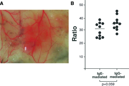
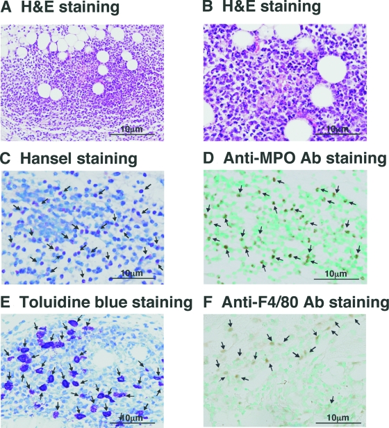
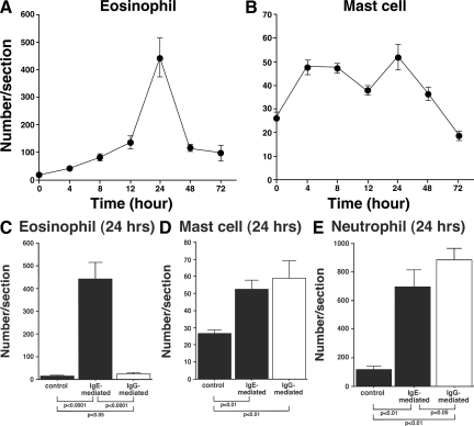
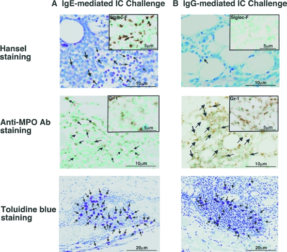
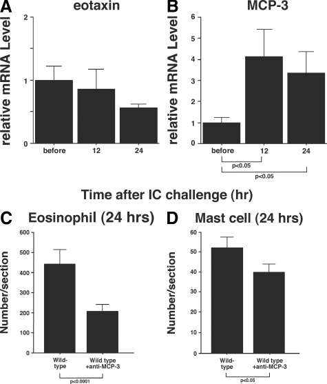
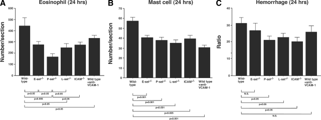
Prominent eosinophil infiltration is a characteristic of some forms of vasculitis, such as Churg-Strauss syndrome, also known as allergic granulomatous vasculitis. In the current study, we established a mouse model of cutaneous eosinophilic vasculitis by the cutaneous reverse passive Arthus reaction using IgE injection instead of IgG. Wild-type C57BL/6 mice were injected with IgE anti-trinitrophenyl antibodies, followed immediately by intravenous administration of trinitrophenyl bovine serum albumin. IgE-mediated immune complex challenge induced substantial hemorrhage with marked infiltration of eosinophils in which neutrophils, mast cells, and macrophages were also mixed. This finding contrasted remarkably with the neutrophil-dominant infiltration pattern in IgG-mediated immune complex challenge. In the lesion, the expression level of monocyte chemotactic protein-3 was increased, and anti-monocyte chemotactic protein-3 treatment resulted in a significant but incomplete blockade of eosinophil recruitment. Furthermore, mice lacking E-selectin, P-selectin, L-selectin, or intercellular adhesion molecule-1, as well as wild-type mice that received anti-vascular cell adhesion molecule-1-blocking antibodies were assessed for the IgE-mediated Arthus reaction. After 24 hours, the loss of P-selectin resulted in a significant reduction in eosinophil accumulation compared with both wild-type mice and other mouse mutants. Collectively, the Fc class of immunoglobulins, which forms these immune complexes, critically determines the disease manifestation of vasculitis. The IgE-mediated cutaneous reverse passive Arthus reaction may serve as an experimental model for cutaneous eosinophilic infiltration in vasculitis as well as in other diseases.
Figures
Similar articles
-
Basophils and mast cells play critical roles for leukocyte recruitment in IgE-mediated cutaneous reverse passive Arthus reaction.J Dermatol Sci. 2012 Sep;67(3):181-9. doi: 10.1016/j.jdermsci.2012.06.005. Epub 2012 Jun 23. J Dermatol Sci. 2012. PMID: 22784785
-
The cutaneous reverse Arthus reaction requires intercellular adhesion molecule 1 and L-selectin expression.J Immunol. 2002 Mar 15;168(6):2970-8. doi: 10.4049/jimmunol.168.6.2970. J Immunol. 2002. PMID: 11884469
-
P-selectin glycoprotein ligand-1 is required for the development of cutaneous vasculitis induced by immune complex deposition.J Leukoc Biol. 2004 Aug;76(2):374-82. doi: 10.1189/jlb.1203650. Epub 2004 May 3. J Leukoc Biol. 2004. PMID: 15123773
-
Role of IgE in the development of allergic airway inflammation and airway hyperresponsiveness--a murine model.Allergy. 1999 Apr;54(4):297-305. doi: 10.1034/j.1398-9995.1999.00085.x. Allergy. 1999. PMID: 10371087 Review.
-
Systemic vasculitis with asthma and eosinophilia: a clinical approach to the Churg-Strauss syndrome.Medicine (Baltimore). 1984 Mar;63(2):65-81. doi: 10.1097/00005792-198403000-00001. Medicine (Baltimore). 1984. PMID: 6366453 Review.
Cited by
-
Mast cell and autoimmune diseases.Mediators Inflamm. 2015;2015:246126. doi: 10.1155/2015/246126. Epub 2015 Apr 5. Mediators Inflamm. 2015. PMID: 25944979 Free PMC article. Review.
-
Mast cell function: a new vision of an old cell.J Histochem Cytochem. 2014 Oct;62(10):698-738. doi: 10.1369/0022155414545334. Epub 2014 Jul 25. J Histochem Cytochem. 2014. PMID: 25062998 Free PMC article. Review.
-
Mast Cells and Interleukins.Int J Mol Sci. 2022 Nov 13;23(22):14004. doi: 10.3390/ijms232214004. Int J Mol Sci. 2022. PMID: 36430483 Free PMC article. Review.
-
Eosinophils in vasculitis: characteristics and roles in pathogenesis.Nat Rev Rheumatol. 2014 Aug;10(8):474-83. doi: 10.1038/nrrheum.2014.98. Epub 2014 Jul 8. Nat Rev Rheumatol. 2014. PMID: 25003763 Free PMC article. Review.
-
Sex Differences in Mast Cell-Associated Disorders: A Life Span Perspective.Cold Spring Harb Perspect Biol. 2022 Oct 3;14(10):a039172. doi: 10.1101/cshperspect.a039172. Cold Spring Harb Perspect Biol. 2022. PMID: 35817512 Free PMC article. Review.
References
-
- Rothenberg ME, Hogan SP. The eosinophil. Annu Rev Immunol. 2006;24:147–174. - PubMed
-
- Silberstein DS. Eosinophil function in health and disease. Crit Rev Oncol Hematol. 1995;19:47–77. - PubMed
-
- Churg J. Allergic granulomatosis and granulomatous-vascular syndromes. Ann Allergy. 1963;21:619–628. - PubMed
-
- Fujimoto M, Sato S, Hayashi N, Wakugawa M, Tsuchida T, Tamaki K. Juvenile temporal arteritis with eosinophilia: a distinct clinicopathological entity. Dermatology. 1996;192:32–35. - PubMed
-
- Chumbley LC, Harrison EG, Jr, DeRemee RA. Allergic granulomatosis and angiitis (Churg-Strauss syndrome). Report and analysis of 30 cases. Mayo Clin Proc. 1977;52:477–484. - PubMed
Publication types
MeSH terms
Substances
LinkOut - more resources
Full Text Sources
Other Literature Sources
Medical
Molecular Biology Databases
