Activation of the endoplasmic reticulum stress-associated transcription factor x box-binding protein-1 occurs in a subset of normal germinal-center B cells and in aggressive B-cell lymphomas with prognostic implications
- PMID: 19389935
- PMCID: PMC2684197
- DOI: 10.2353/ajpath.2009.080848
Activation of the endoplasmic reticulum stress-associated transcription factor x box-binding protein-1 occurs in a subset of normal germinal-center B cells and in aggressive B-cell lymphomas with prognostic implications
Abstract
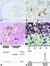
X box-binding protein 1 (Xbp-1) is a transcription factor that is required for the terminal differentiation of B lymphocytes into plasma cells. The Xbp-1 gene is activated in response to endoplasmic reticulum stress signals, which generate a 50-kDa nuclear protein that acts as a potent transactivator and regulates the expression of genes related to the unfolded protein response. Activated Xbp-1 is essential for cell survival in plasma-cell tumors but its role in B-cell lymphomas is unknown. We analyzed the expression of activated Xbp-1 in reactive lymphoid tissues, 411 lymphomas and plasma-cell neoplasms, and 24 B-cell lines. In reactive tissues, Xbp-1 was only found in nuclear extracts. Nuclear expression of Xbp-1 was observed in occasional reactive plasma cells and in a subpopulation of Irf-4(+)/Bcl-6(-)/Pax-5(-) B cells in the light zones of reactive germinal centers, probably representing cells committed to plasma-cell differentiation. None of the low-grade lymphomas showed evidence of Xbp-1 activation; however, Xbp-1 activation was found in 28% of diffuse large B-cell lymphomas, independent of germinal or postgerminal center phenotype, as well as in 48% of plasmablastic lymphomas and 69% of plasma-cell neoplasms. Diffuse large B-cell lymphomas with nuclear Xbp-1 expression had a significantly worse response to therapy and shorter overall survival compared with negative tumors. These findings suggest that Xbp-1 activation may play a role in the pathogenesis of aggressive B-cell lymphomas.
Figures


Similar articles
-
Expression of the interferon regulatory factor 8/ICSBP-1 in human reactive lymphoid tissues and B-cell lymphomas: a novel germinal center marker.Am J Surg Pathol. 2008 Aug;32(8):1190-200. doi: 10.1097/PAS.0b013e318166f46a. Am J Surg Pathol. 2008. PMID: 18580679 Free PMC article.
-
Tissue inhibitor of metalloproteinase 1 (TIMP-1) promotes plasmablastic differentiation of a Burkitt lymphoma cell line: implications in the pathogenesis of plasmacytic/plasmablastic tumors.Blood. 2005 Feb 15;105(4):1660-8. doi: 10.1182/blood-2004-04-1385. Epub 2004 Oct 12. Blood. 2005. PMID: 15479729
-
Expression pattern of XBP1(S) in human B-cell lymphomas.Haematologica. 2009 Mar;94(3):419-22. doi: 10.3324/haematol.2008.001156. Epub 2009 Jan 27. Haematologica. 2009. PMID: 19176362 Free PMC article.
-
The X-box binding protein-1 transcription factor is required for plasma cell differentiation and the unfolded protein response.Immunol Rev. 2003 Aug;194:29-38. doi: 10.1034/j.1600-065x.2003.00057.x. Immunol Rev. 2003. PMID: 12846805 Review.
-
Expression of apoptosis regulators in germinal centers and germinal center-derived B-cell lymphomas: insight into B-cell lymphomagenesis.Pathol Int. 2007 Jul;57(7):391-7. doi: 10.1111/j.1440-1827.2007.02115.x. Pathol Int. 2007. PMID: 17587238 Review.
Cited by
-
Expression of peroxiredoxins I and IV in multiple myeloma: association with immunoglobulin accumulation.Virchows Arch. 2013 Jul;463(1):47-55. doi: 10.1007/s00428-013-1433-1. Epub 2013 Jun 5. Virchows Arch. 2013. PMID: 23737084
-
Induction of Kaposi's Sarcoma-Associated Herpesvirus-Encoded Viral Interleukin-6 by X-Box Binding Protein 1.J Virol. 2015 Oct 21;90(1):368-78. doi: 10.1128/JVI.01192-15. Print 2016 Jan 1. J Virol. 2015. PMID: 26491160 Free PMC article.
-
Aggressive large B-cell lymphoma with plasma cell differentiation: immunohistochemical characterization of plasmablastic lymphoma and diffuse large B-cell lymphoma with partial plasmablastic phenotype.Haematologica. 2010 Aug;95(8):1342-9. doi: 10.3324/haematol.2009.016113. Epub 2010 Apr 23. Haematologica. 2010. PMID: 20418245 Free PMC article.
-
ALS-associated TDP-43 induces endoplasmic reticulum stress, which drives cytoplasmic TDP-43 accumulation and stress granule formation.PLoS One. 2013 Nov 29;8(11):e81170. doi: 10.1371/journal.pone.0081170. eCollection 2013. PLoS One. 2013. PMID: 24312274 Free PMC article.
-
Correlation Between Low Cytoplasmic Expression of XBP1 and the Likelihood of Surviving Hepatocellular Carcinoma.In Vivo. 2024 May-Jun;38(3):1316-1324. doi: 10.21873/invivo.13571. In Vivo. 2024. PMID: 38688649 Free PMC article.
References
-
- Brodsky JL, McCracken AA. ER protein quality control and proteasome-mediated protein degradation. Sem Cell Dev Biol. 1999;10:507–513. - PubMed
-
- Rutkowski DT, Kaufman RJ. A trip to the ER: coping with stress. Trends Cell Biol. 2004;14:20–28. - PubMed
-
- Haze K, Okada T, Yoshida H, Yanagi H, Yura T, Negishi M, Mori K. Identification of the G13 (cAMP-response-element-binding protein-related protein) gene product related to activating transcription factor 6 as a transcriptional activator of the mammalian unfolded protein response. Biochem J. 2001;355:19–28. - PMC - PubMed
Publication types
MeSH terms
Substances
LinkOut - more resources
Full Text Sources
Research Materials

