. 2009 Apr;5(4):e1000397.
doi: 10.1371/journal.ppat.1000397. Epub 2009 Apr 24.

Mechanism of genomic instability in cells infected with the high-risk human papillomaviruses

Affiliations

Mechanism of genomic instability in cells infected with the high-risk human papillomaviruses

Meelis Kadaja et al. PLoS Pathog. 2009 Apr.

Abstract

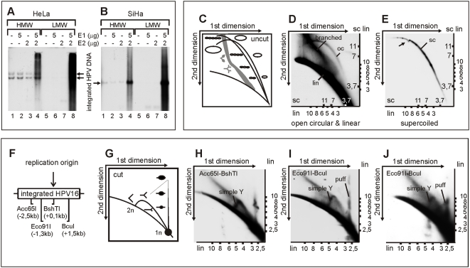
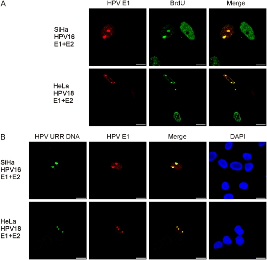
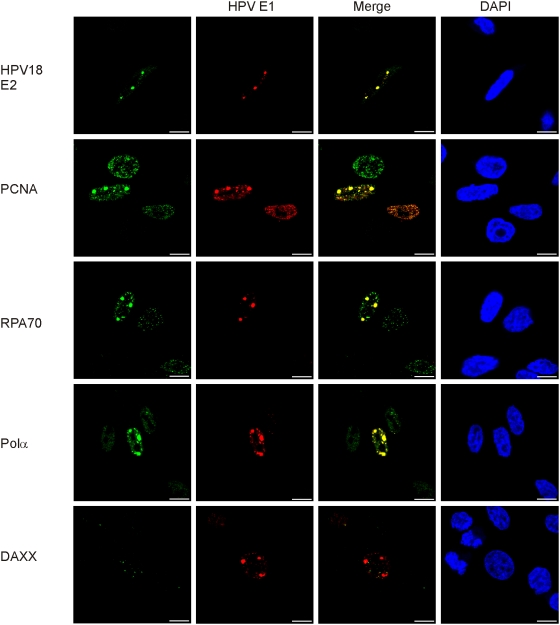
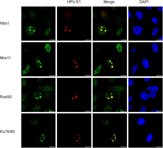
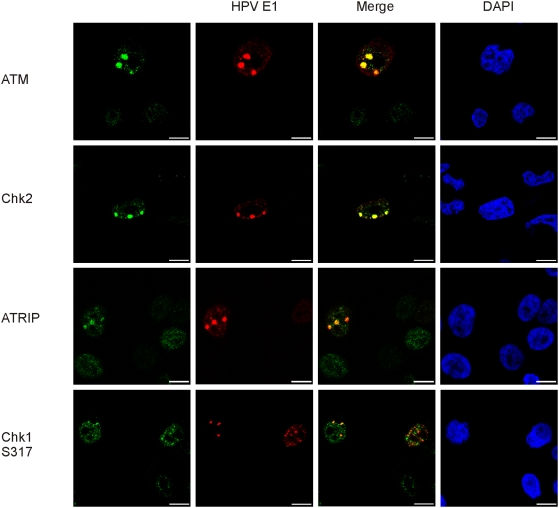
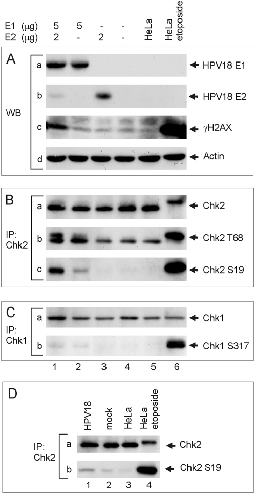
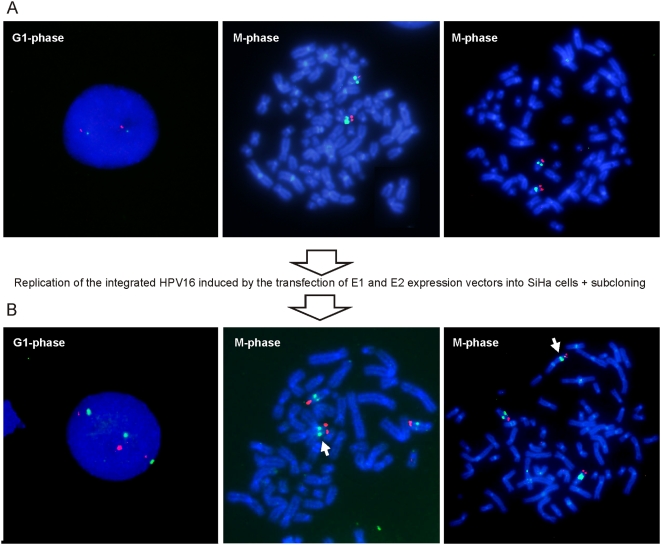
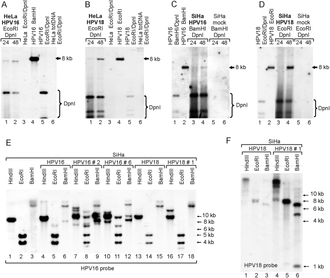
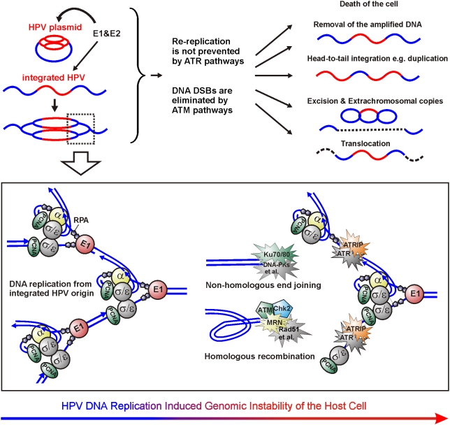
In HPV-related cancers, the "high-risk" human papillomaviruses (HPVs) are frequently found integrated into the cellular genome. The integrated subgenomic HPV fragments express viral oncoproteins and carry an origin of DNA replication that is capable of initiating bidirectional DNA re-replication in the presence of HPV replication proteins E1 and E2, which ultimately leads to rearrangements within the locus of the integrated viral DNA. The current study indicates that the E1- and E2-dependent DNA replication from the integrated HPV origin follows the "onion skin"-type replication mode and generates a heterogeneous population of replication intermediates. These include linear, branched, open circular, and supercoiled plasmids, as identified by two-dimensional neutral-neutral gel-electrophoresis. We used immunofluorescence analysis to show that the DNA repair/recombination centers are assembled at the sites of the integrated HPV replication. These centers recruit viral and cellular replication proteins, the MRE complex, Ku70/80, ATM, Chk2, and, to some extent, ATRIP and Chk1 (S317). In addition, the synthesis of histone gammaH2AX, which is a hallmark of DNA double strand breaks, is induced, and Chk2 is activated by phosphorylation in the HPV-replicating cells. These changes suggest that the integrated HPV replication intermediates are processed by the activated cellular DNA repair/recombination machinery, which results in cross-chromosomal translocations as detected by metaphase FISH. We also confirmed that the replicating HPV episomes that expressed the physiological levels of viral replication proteins could induce genomic instability in the cells with integrated HPV. We conclude that the HPV replication origin within the host chromosome is one of the key factors that triggers the development of HPV-associated cancers. It could be used as a starting point for the "onion skin"-type of DNA replication whenever the HPV plasmid exists in the same cell, which endangers the host genomic integrity during the initial integration and after the de novo infection.


Conflict of interest statement

The authors have declared that no competing interests exist.

Figures

Figure 1
Figure 1. DNA replication initiated from the integrated HPV origin generates various low-molecular-weight DNA products.
High-molecular-weight (HMW) DNA and low-molecular-weight (LMW) DNA were fractionated and purified by Hirt lysis 24 hrs post-transfection from HeLa cells (A) and from SiHa cells (B) that were both transfected as follows: mock-transfection (lanes 1 and 5); 5 µg of HPV18 E1 expression plasmid (lanes 2 and 6); 2 µg of E2 expression plasmid (lanes 3 and 7); 5 µg of HPV18 E1 and 2 µg of E2 expression plasmids (lanes 4 and 8). A 3 µg portion of HMW DNA and three times the respective amount of LMW DNA were digested with HindIII (A) or Acc65I/BshTI (B) and separated on a one-dimensional gel. The integrated HPV URR-specific signals were detected by Southern blot analysis. (C) Schematic presentation of the migration of dsDNA linear, supercoiled, and open circle molecules on 2D neutral-neutral gels. (D, E) 5 µg of HPV18 E1 and 2 µg of E2 expression plasmids were transfected into HeLa cells and LMW DNA was purified by Hirt lysis 48 hrs post-transfection. Extracted DNA was digested with DpnI and fractionated by the CsCl-ethidium bromide density gradient. The fraction of linear fragments (lin) and open circular molecules (oc) (D) and the fraction of supercoiled circular plasmids (sc) (E) were separated on a 2D gel, transferred to a nylon filter, and probed with the HPV18 genomic fragment (from nt 3917 to1575). Numbers shown on the axes represent the markers of linear (lin) and supercoiled circular (sc) DNA forms. Black arrowhead indicates the shift that was caused by mtDNA. (F) Schematic presentation of HPV16 integration locus within chromosome 13 in SiHa cells, where the cleavage sites of Acc65I, Eco91I, BshTI, and BcuI as well as their distances from HPV origin are presented. (G) Schematic presentation of the migration of replication forks and replication puffs on 2D neutral-neutral gels. (H,I) SiHa cells were co-transfected with 5 µg of HPV18 E1 and 2 µg of E2 expression plasmids. LMW DNA was extracted 24 hrs post-transfection and digested with Acc65I-BshTI (H) and Eco91I-BcuI (I). Respective HPV16 genome fragments were used as probes on 2D Southern blots. (J) SiHa cells were co-transfected with 1 µg of HPV18 E1 and 2 µg of E2 expression plasmids. Extracted LMW DNA was digested with Eco91I-BcuI and analyzed by 2D Southern blots.
Figure 2
Figure 2. DNA replication of the integrated HPV takes place at specific nuclear foci.
HeLa and SiHa cells were co-transfected with 5 µg of HPV18 E1 and 2 µg of E2 expression plasmids. Cells were analyzed 20 hrs post-transfection. (A) Co-immunostaining of E1 (Alexa Fluor 568, first column) and BrdU (FITC, second column). Cells were pulse-labeled with BrdU for 2 hrs prior to IF analysis. In the third column, the localizations of E1 and BrdU in the same cells are presented as a merged image. (B) Combined immunofluorescence and FISH analysis to detect the integrated HPV DNA (Alexa Fluor 488, first column) and the HPV E1 protein (Alexa Fluor 568, second column) in SiHa and HeLa cells. Localizations of the E1 and the integrated HPV DNA are also presented in the third column as a merged image. DNA was counterstained with DAPI (fourth column).
Figure 3
Figure 3. Cellular replication factors colocalize with the HPV 18 E1 in HeLa cells.
Cells were transfected and analyzed as described previously. Co-immunostaining of HPV18 E1 (Alexa Fluor 568, second column) together with the following proteins are shown: HPV18 E2, PCNA, RPA, polymerase α/primase, and Daxx (Alexa Fluor 488, first column). Merged images are presented in the third column and DAPI stained nuclei in the fourth column.
Figure 4
Figure 4. Replication centers of the integrated HPV recruit Mre11-Nbs1-Rad50 complex and Ku70/80 heterodimer.
HeLa cells were transfected and analyzed as described previously. Co-immunostaining of HPV18 E1 (Alexa Fluor 568, second column) and the following proteins are presented: Mre11, Nbs1, Rad50, and Ku70/80 proteins (Alexa Fluor 488, first column). The localizations of the E1 and the respective DNA repair proteins are also shown in the third column as a merged image and DAPI stained nuclei are presented in the fourth column.
Figure 5
Figure 5. Repair of the amplified HPV18 integration locus is coordinated by the ATM and ATR signaling pathways.
HeLa cells were transfected and analyzed as described previously. Co-immunostaining of HPV18 E1 (Alexa Fluor 568, second column) and the following proteins are shown at the figure: ATM, Chk2, ATRIP, and Chk1 (S317) (Alexa Fluor 488, first column). Merged images are shown in the third column and DAPI stained nuclei in the fourth column.
Figure 6
Figure 6. Activation of the ATM-Chk2 signaling pathway.
(A–C) HeLa cells were transfected as follows: 5 µg of HPV18 E1 and 2 µg of HPV18 E2 expression plasmids (lane 1); 5 µg of HPV18 E1 expression plasmid (lane 2); 2 µg of E2 expression plasmid (lane 3); and a mock-transfection (lane 4). In every transfection, the amount of plasmid was adjusted to 10 µg with a carrier plasmid (pauxoMCF). Non-transfected HeLa cells are presented in lane 5 and HeLa cells that were treated 1 h with etoposide (50 µM) prior to the analysis in lane 6. Western blot analyses were performed at a 24 hrs time point to detect HPV18 E1 (A, panel a), HPV18 E2 (A, panel b), gamma histone H2AX (phosphorylated at S139) (A, panel c), and β-actin (A, panel d). Western blot analyses of Chk2 phosphorylated at Thr68 and Ser19 were performed after the immunoprecipitation with the anti-Chk2 antibody (B, panels a, b, c). Chk1 phosphorylated at Ser317 was detected from extracts that were immunoprecipitated with the anti-Chk1 antibody (C, panels a, b). (D) HeLa cells were transfected either with 2 µg of circular HPV18 genome, 2 µg of pBabePuro and 6 µg of carrier plasmid (lane 1), or with 2 µg of pBabePuro and 8 µg of carrier plasmid (lane 2). Untransfected cells were removed with puromycin treatment (2 µg/ml) 24–48 h posttransfection. Western blot analyses with anti-Chk2 (panel a) and anti-Chk2-Ser19 (panel b) antibodies were performed after the immunoprecipitation with the anti-Chk2 antibody at a 72 h time point. Untreated HeLa cells are shown in lane 3 and etoposide-treated HeLa cells in lane 4.
Figure 7
Figure 7. Re-replication of the integrated HPV induces the instability of chromosome structure.
SiHa cells were transfected with 10 µg of HPV18 E1 and 5 µg of E2 expression plasmids. Single cell subcloning was performed 72 h after transfection . SiHa cells and the subclones with novel restriction patterns were analyzed by FISH. (A) FISH analysis of the interphase nuclei (left panel) and metaphase chromosome spreads (the middle and the right panels) of SiHa cells. (B) Interphase FISH (left panel) and metaphase FISH (the middle and the right panels) analyses of the subclone with cross-chromosomal translocation. The integrated HPV16 is visible as green (Alexa Fluor 488), and the subtelomeric region of the chromosome 13 is visible as red (Texas Red). The translocation of 13q is indicated with the white arrowhead.
Figure 8
Figure 8. The HPV genome induces genomic instability in cells harboring the integrated HPV.
Southern blot analyses of the HeLa (A, B) and SiHa cells (C, D) co-transfected with 1 µg of HPV16 genome and 1 µg of linearized pEGFPN-1 (A, C) or with 1 µg of HPV18 genome and 1 µg of linearized pEGFPN-1 (B, D). Low-molecular-weight DNA was extracted 24 h and 48 h after transfection and digested with restriction enzymes, as indicated in the figure. HPV plasmids were detected with a 32P-labeled HPV16 or HPV18 genome probe, respectively. (E, F) The restriction analyses of untreated SiHa cells (E, lanes 1–3), HPV16-transfected SiHa cells five weeks post-transfection (E, lanes 4–6), HPV18-transfected SiHa cells five weeks post-transfection (E, lanes 13–15; F, lanes 1–3), as well as the subclones of the HPV16-transfected SiHa cells (E, lanes 7–12) and HPV18-transfected SiHa cells (E, lanes 16–18; F, lanes 4–6). Digested total DNA was analyzed by Southern blotting, where the HPV16 (E) or HPV18 (F) genomes were used as probes. Restriction enzymes are indicated in the figure.
Figure 9
Figure 9. Mechanism of the genomic instability of the cells harboring the replication origin of integrated HPV.
If HPV plasmid is present in the cells harboring integrated HPV, the DNA re-replication from the integrated HPV origin is initiated, and ATM and ATR signaling pathways respond, respectively, to the produced DSBs and ssDNA. In most cases, the cells will either become apoptotic or the damage sites will be properly repaired by homologous recombination and non-homologous end-joining. In addition, duplications within the HPV locus have previously been detected. In the current study, the excision and generation of extrachromosomal copies of the HPV locus and cross-chromosomal translocations were also detected. All these mutations require DSBs that could be generated, such as through head-to-tail fork collision or by encounter of the re-replication fork with the Okazaki fragments of the previous fork. Supported by our observations, we speculate that the cellular defense against the integrated HPV re-replication is primary coordinated by ATM. ATR pathways do not interfere with a properly working E1-driven replication fork but, rather, are responsible for the repair of the damage that is caused by fork collision and dissociation that reveals the RPA coated ssDNA without the generation of DSBs. DNA replication/repair factors that are shown to be within the HPV replication centers in the current work, are colored in the figure.

References

    1. Howley PM, Lowy DR. Papillomaviruses and Their Replication. In: Knipe DM, aH PM, editors. Philadelphia: Lippincott Williams & Wilkins; 2001. pp. 2197–2230.
    1. Ustav M, Stenlund A. Transient replication of BPV-1 requires two viral polypeptides encoded by the E1 and E2 open reading frames. Embo J. 1991;10:449–457. - PMC - PubMed
    1. Chiang CM, Ustav M, Stenlund A, Ho TF, Broker TR, et al. Viral E1 and E2 proteins support replication of homologous and heterologous papillomaviral origins. Proc Natl Acad Sci U S A. 1992;89:5799–5803. - PMC - PubMed
    1. Sverdrup F, Khan SA. Replication of human papillomavirus (HPV) DNAs supported by the HPV type 18 E1 and E2 proteins. J Virol. 1994;68:505–509. - PMC - PubMed
    1. Kadaja M, Silla T, Ustav E, Ustav M. Papillomavirus DNA replication - from initiation to genomic instability. Virology. 2009;384:360–368. - PubMed

Publication types

Substances