. 2009 Apr;5(4):e1000396.
doi: 10.1371/journal.ppat.1000396. Epub 2009 Apr 24.

Naturally occurring lipid A mutants in neisseria meningitidis from patients with invasive meningococcal disease are associated with reduced coagulopathy

Affiliations

Naturally occurring lipid A mutants in neisseria meningitidis from patients with invasive meningococcal disease are associated with reduced coagulopathy

Floris Fransen et al. PLoS Pathog. 2009 Apr.

Abstract

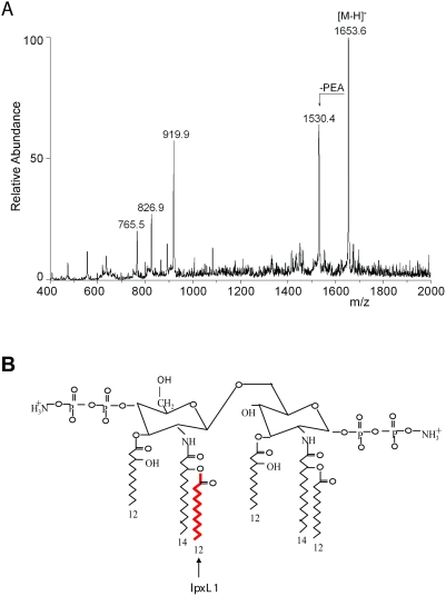
Neisseria meningitidis is a major cause of bacterial meningitis and sepsis worldwide. Lipopolysaccharide (LPS), a major component of the Gram-negative bacterial outer membrane, is sensed by mammalian cells through Toll-like receptor 4 (TLR4), resulting in activation of proinflammatory cytokine pathways. TLR4 recognizes the lipid A moiety of the LPS molecule, and the chemical composition of the lipid A determines how well it is recognized by TLR4. N. meningitidis has been reported to produce lipid A with six acyl chains, the optimal number for TLR4 recognition. Indeed, meningococcal sepsis is generally seen as the prototypical endotoxin-mediated disease. In the present study, we screened meningococcal disease isolates from 464 patients for their ability to induce cytokine production in vitro. We found that around 9% of them were dramatically less potent than wild-type strains. Analysis of the lipid A of several of the low-activity strains by mass spectrometry revealed they were penta-acylated, suggesting a mutation in the lpxL1 or lpxL2 genes required for addition of secondary acyl chains. Sequencing of these genes showed that all the low activity strains had mutations that inactivated the lpxL1 gene. In order to see whether lpxL1 mutants might give a different clinical picture, we investigated the clinical correlate of these mutations in a prospective nationwide observational cohort study of adults with meningococcal meningitis. Patients infected with an lpxL1 mutant presented significantly less frequently with rash and had higher thrombocyte counts, consistent with reduced cytokine induction and less activation of tissue-factor mediated coagulopathy. In conclusion, here we report for the first time that a surprisingly large fraction of meningococcal clinical isolates have LPS with underacylated lipid A due to mutations in the lpxL1 gene. The resulting low-activity LPS may have an important role in virulence by aiding the bacteria to evade the innate immune system. Our results provide the first example of a specific mutation in N. meningitidis that can be correlated with the clinical course of meningococcal disease.


Conflict of interest statement

The authors have declared that no competing interests exist.

Figures

Figure 1
Figure 1. Strain HF13 is an lpxL1 mutant.
(A) Mass spectrum of HF13 lipid A. The highest peak (1653.6) corresponds to penta-acylated lipid A with two phosphate groups and one phosphoethanolamine (PEA); the second peak (1530.4) corresponds to penta-acytlated lipid A with two phosphate groups without PEA. (B) Depiction of N. meningitidis wild-type lipid A. The acyl chain that is added by LpxL1 is indicated.
Figure 2
Figure 2. Presentation of all lpxL1 mutations.
The lpxL1 gene sequence of strain MC58 is shown including the different types of mutations and their positions in the gene found among isolates from patients. Each type of mutation is indicated with a different color. An ‘_’ indicates a nucleotide that is deleted in the mutant strain, pink indicates an insertion element (type I and II mutations), yellow indicates a deletion of a guanine in a stretch of five guanines (type III mutation), green indicates a deletion of an adenosine in a stretch of five adenosines (type IV mutation), light blue indicates a deletion of an adenosine in a stretch of seven adenosines (type V mutation), dark blue indicates an insertion of an adenosine in a stretch of seven adenosines (type V mutation), and the sequences that are highlighted in red or gray are deleted in the mutant strain (type VI and VII mutations respectively).
Figure 3
Figure 3. Comparison of IL-6 induction in MM-6 cells between wild-type strains and lpxL1 mutants.
MM-6 cells were stimulated for 18 h with titrations of indicated strains and IL-6 in supernatant was quantified with ELISA. H44/76 is a wild-type strain, L8 lpxL1 is a constructed lpxL1 mutant, pLAK33 is an LPS-deficient mutant, and all other strains are spontaneous lpxL1 mutants. Results of one representative experiment of three independent experiments are shown. Error bars indicate S.E.M. of triplicates.
Figure 4
Figure 4. Comparison between wild-type strains and lpxL1 mutants of IL-8 induction in HEK293 cells transfected with human TLR4.
HEK293 cells transfected with human TLR4, CD14, and MD-2 were stimulated for 18 h with titrations of indicated strains and IL-8 in supernatant was quantified with ELISA. H44/76 is a wild-type strain, L8 lpxL1 is a constructed lpxL1 mutant, pLAK33 is an LPS-deficient mutant, and all other strains are spontaneous lpxL1 mutants. Results of one representative experiment of three independent experiments are shown. Error bars indicate S.E.M. of triplicates.
Figure 5
Figure 5. Comparison between wild-type strains and lpxL1 mutants in pro-inflammatory cytokine induction in PBMCs.
PBMCs from three different donors were stimulated with titrations of the indicated strains and IL-6, TNF-α, and IL-1β were quantified in the supernatant 18 h after stimulation. H44/76 is a wild-type strain, L8 lpxL1 is a constructed lpxL1 mutant, pLAK33 is an LPS-deficient mutant, and all other strains are spontaneous lpxL1 mutants. Results of one representative experiment of two independent experiments are shown. Error bars indicate S.E.M. of triplicates.
Figure 6
Figure 6. Clinical correlate of lpxL1 mutations in meningococcal meningitis.
(A) Frequency of rash in patients presenting with meningitis infected by lpxL1 wild-type and mutant strains. (B,C) Platelet counts on admission for lpxL1 wild-type and mutant strains (B) and patients presenting with and without rash (C). Horizontal bars reflect medians. The Mann-Whitney U test was used to identify differences between groups in continuous variables, and dichotomous variables were compared by the chi-square or Fisher exact test.

References

    1. Stephens DS, Greenwood B, Brandtzaeg P. Epidemic meningitis, meningococcaemia, and Neisseria meningitidis. Lancet. 2007;369:2196–2210. - PubMed
    1. Beutler B, Rietschel ET. Innate immune sensing and its roots: the story of endotoxin. Nat Rev Immunol. 2003;3:169–176. - PubMed
    1. Palsson-McDermott EM, O'Neill LA. Signal transduction by the lipopolysaccharide receptor, Toll-like receptor-4. Immunology. 2004;113:153–162. - PMC - PubMed
    1. Parrillo JE. Pathogenetic mechanisms of septic shock. N Engl J Med. 1993;328:1471–1477. - PubMed
    1. Russell JA. Management of sepsis. N Engl J Med. 2006;355:1699–1713. - PubMed

Publication types