. 2009:2009:526963.
doi: 10.1155/2009/526963. Epub 2009 Apr 14.

Anti-EGFR Therapy: Mechanism and Advances in Clinical Efficacy in Breast Cancer

Affiliations

Anti-EGFR Therapy: Mechanism and Advances in Clinical Efficacy in Breast Cancer

John F Flynn et al. J Oncol. 2009.

Abstract

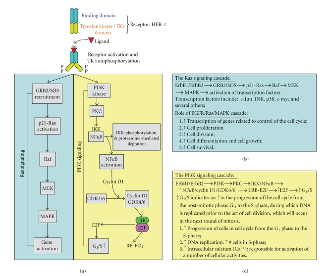
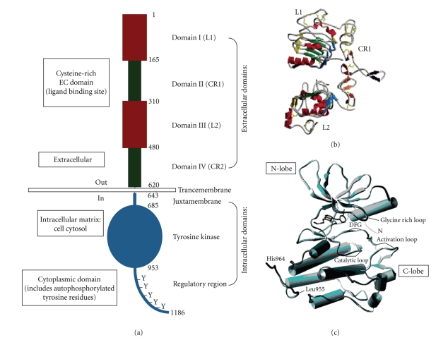
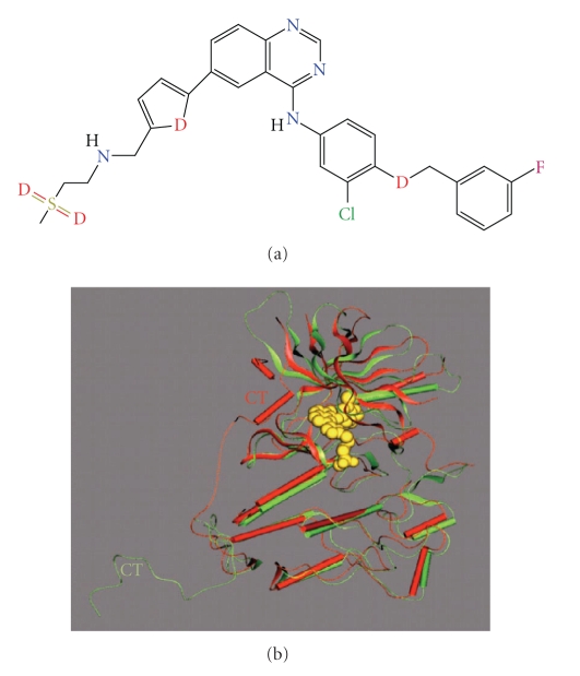
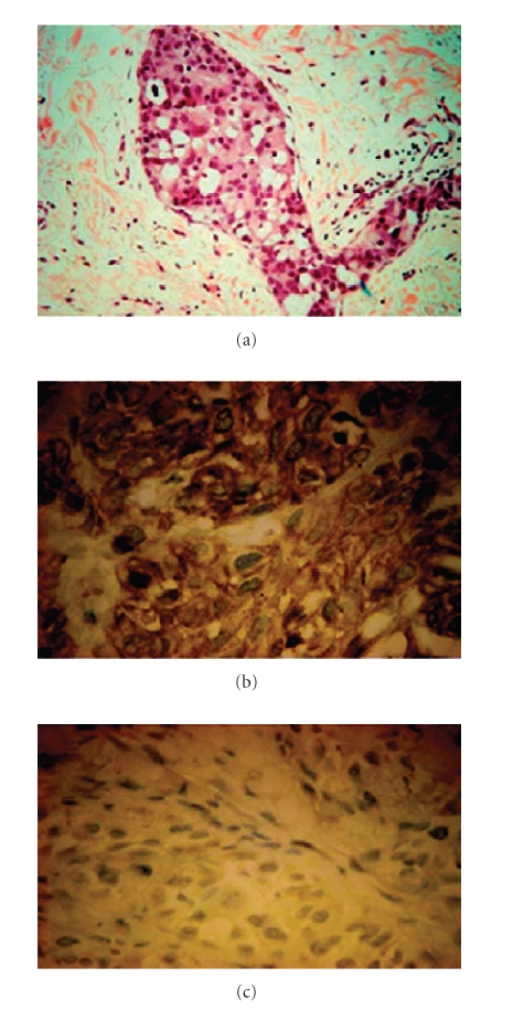
This review will focus on recent advances in the application of antiepidermal growth factor receptor (anti-EGFR) for the treatment of breast cancer. The choice of EGFR, a member of the ErbB tyrosine kinase receptor family, stems from evidence pinpointing its role in various anti-EGFR therapies. Therefore, an increase in our understanding of EGFR mechanism and signaling might reveal novel targets amenable to intervention in the clinic. This knowledge base might also improve existing medical treatment options and identify research gaps in the design of new therapeutic agents. While the approved use of drugs like the dual kinase inhibitor Lapatinib represents significant advances in the clinical management of breast cancer, confirmatory studies must be considered to foster the use of anti-EGFR therapies including safety, pharmacokinetics, and clinical efficacy.


Figures

Figure 1
Figure 1
The EGFR Signaling Pathway. (a) Upon EGF-ligand binding to the EGFR there is subsequent dimerization (homo- or hetero-) and tyrosine kinase residue auto-/transphosphorylation of dimer partners, which in turn initiates the actual downstream signaling pathways. (b) Ras signaling cascade in tabulated form. (c) PI3K signaling cascade in tabulated form.
Figure 2
Figure 2
(a) Basic Structure of EGFR demonstrating relevant domains. (I) The extracellular domains: (1) domain I: L1; (2) domain II: CR1; domain III: L2; domain IV: CR2. (II) Transmembrane domains. (III) The intracellular domains (1) juxtamembrane domain; (2) tyrosine kinase domain; (3) regulatory region domain. The phosphorylation of several substrates by the tyrosine kinase domain of the EGFR receptor is responsible for activating the various signaling cascades seen in Figure 1. (b) Structure of domains I–IV of EGFR (no ligand bound). Note the “protruding loop” in domain II (CR1) directed away from the C-shaped region of the ligand-binding zone formed by domains I, II, and III. (c) The tyrosine kinase domain of EGFR showing the N-lobe and C-lobe flanking the activation loop and active site cleft [2, 3].
Figure 3
Figure 3
Molecular and crystal structures of EGFR inhibitor Lapatinib and Lapatinib bound and complexed to EGFR ATP-binding pocket, respectively. (a) Molecular structure of Lapatinib (CID208908), an EGFR-ErbB2 inhibitor. (b) Overlay of EGFR in the Lapatinib and Erlotinib complexes. EGFR in the Lapatinib and Erlotinib structures is shown as red and green ribbons, respectively. Lapatinib is shown as a yellow space-filling model. The two proteins were overlaid based on residues in the COOH-terminal domain of the kinase. The COOH-terminal in both structures is CT. Disordered residues in the COOH-terminal tail of EGFR are indicated by a dashed line. The figure was prepared using QUANTA (Accelrys), adapted from Wood et al. [36].
Figure 4
Figure 4
Immunohistochemical staining demonstrating the clinical efficacy of Lapatinib. Figure 4 identifies inhibition of activated, phosphorylated ErbB2/HER-2/neu (p-ErbB2) in a breast cancer patient responding to Lapatinib treatment. (a) Shows a dermal-lymphatic invasion (magenta) that is consistent with recurrent inflammatory breast cancer. (b) and (c) Show further immunohistochemical staining for p-ErbB2 performed on tumor biopsy samples obtained from patient X on days 0 (4B) and 21 (4C) of Lapatinib therapy; note the change in positive staining (brownish-yellow). There is a significant decrease in the activation of p-ErbB2 in response to Lapatinib [37, 38].

Similar articles

Cited by

References

    1. Moy B, Goss PE. Lapatinib: current status and future directions in breast cancer. Oncologist. 2006;11(10):1047–1057. - PubMed
    1. Boyd MC. A Literature Review Submitted to the Program for Biochemistry and Molecular Biology at New York Medical College in Partial Fulfillment of the Requirements for the Degree of Master of Science. Valhalla, NY, USA: Laboratory of Dr. Joseph Wu, New York Medical College; 2005. Epidermal growth factor receptor: structural basis for functional properties.
    1. Gotoh N, Tojo A, Hino M, Yazaki Y, Shibuya M. A highly conserved tyrosine residue at codon 845 within the kinase domain is not required for the transforming activity of human epidermal growth factor receptor. Biochemical and Biophysical Research Communications. 1992;186(2):768–774. - PubMed
    1. Konecny GE, Pegram MD, Venkatesan N, et al. Activity of the dual kinase inhibitor lapatinib (GW572016) against HER-2-overexpressing and trastuzumab-treated breast cancer cells. Cancer Research. 2006;66(3):1630–1639. - PubMed
    1. Agrawal A, Gutteridge E, Gee JMW, Nicholson RI, Robertson JFR. Overview of tyrosine kinase inhibitors in clinical breast cancer. Endocrine-Related Cancer. 2005;12(supplement 1):S135–S144. - PubMed