

Review
doi: 10.1007/s00018-009-0026-2.
Epub 2009 Apr 24.
The development and function of regulatory T cells
Affiliations
- PMID: 19390784
- PMCID: PMC2715449
- DOI: 10.1007/s00018-009-0026-2
Item in Clipboard
Review
The development and function of regulatory T cells
Cell Mol Life Sci.
2009 Aug.
Abstract
Regulatory T cells (Tregs) are a critical subset of T cells that mediate peripheral tolerance. There are two types of Tregs: natural Tregs, which develop in the thymus, and induced Tregs, which are derived from naive CD4(+) T cells in the periphery. Tregs utilize a variety of mechanisms to suppress the immune response. While Tregs are critical for the peripheral maintenance of potential autoreactive T cells, they can also be detrimental by preventing effective anti-tumor responses and sterilizing immunity against pathogens. In this review, we will discuss the development of natural and induced Tregs as well as the role of Tregs in a variety of disease settings and the mechanisms they utilize for suppression.
Figures
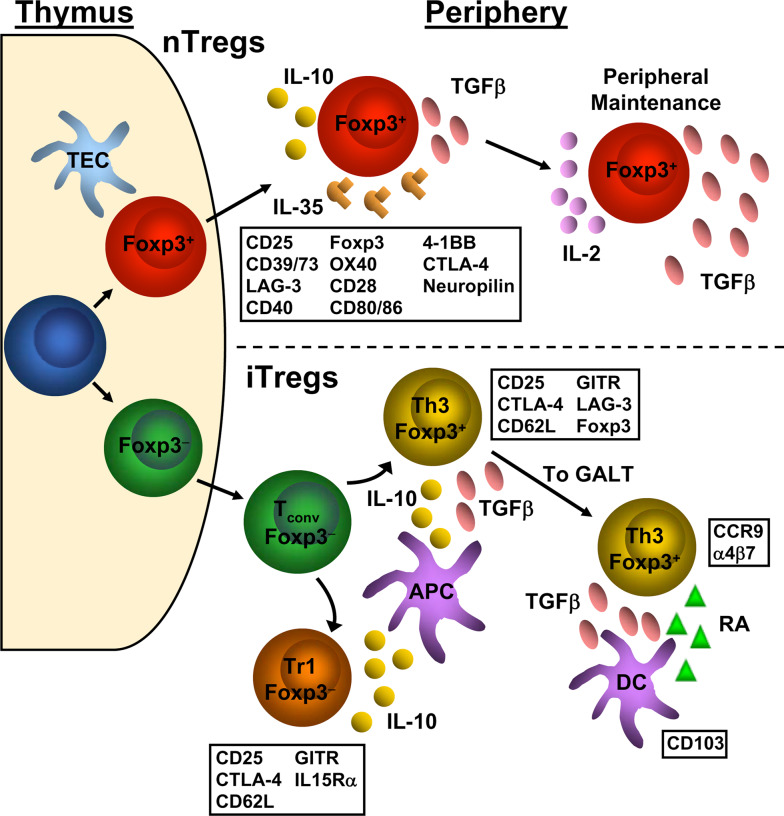
Development of nTregs and iTregs and the relevant markers associated with each. nTregs (top) differentiate from naive conventional T cells to Foxp3+ Tregs in the thymus. In the periphery, natural Tregs express a number of cell surface markers, indicated in the box below the depiction of the natural Treg. However, none of these cell surface markers are unique to Tregs as they are also found on activated conventional T cells. Natural Tregs utilize the cytokines IL-10, IL-35, and TGFβ to exert their suppressive effects upon conventional T cells. TGFβ and IL-2 have also been shown to be important to the maintenance and fidelity of the Treg signature. iTregs (bottom) can be generated from conventional T cell precursors. Once in the periphery, naíve conventional T cells can be induced to become Foxp3− Tr1 cells or Foxp3+ Th3 cells via IL-10 and/or TGFβ secreted by APCs such as dendritic cells and macrophages. These induced Tregs share similar cell surface markers as natural Tregs. Foxp3+-induced Tregs can accumulate in the gut through upregulation of CCR9 and α4β7 via TGFβ and retinoic acid produced by CD103+ dendritic cells. TEC Thymic epithelial cell, T
conv conventional T cell, DC dendritic cell, RA retinoic acid
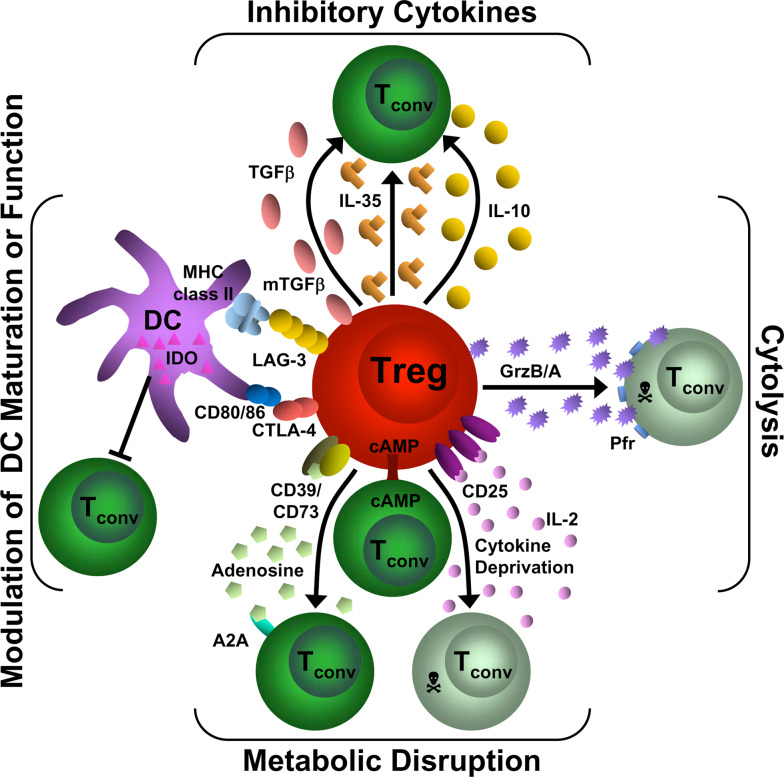
Mechanisms of Treg suppression. This diagram depicts the four basic modes of Treg suppression. A primary mode of Treg suppression is mediated through the inhibitory cytokines IL-10, IL-35, and TGFβ. Tregs also induce cytolysis through granzyme A/B and perforin. They can disrupt metabolic function by IL-2 deprivation which results in apoptosis, cAMP inhibition or by CD39/CD73- generated A2A-mediated immunosuppression. Tregs can also modulate DC maturation or function via a CD80/86 and CTLA-4 interaction or through a LAG-3 and MHC class II interaction. In addition, they can induce the upregulation of IDO in DCs. T
conv Conventional T cell, GrzB/A granzyme B or A, Pfr perforin, cAMP cyclic adenosine monophosphate, A2A adenosine-purinergic adenosine receptor, IDO indoleamine 2,3-dioxygenase, DC dendritic cell
References
-
- Gershon RK. A disquisition on suppressor T cells. Transplant Rev. 1975;26:170–185. - PubMed
-
- Gershon RK, Cohen P, Hencin R, Liebhaber SA. Suppressor T cells. J Immunol. 1972;108:586–590. - PubMed
-
- Sakaguchi S, Sakaguchi N, Asano M, Itoh M, Toda M. Immunologic self-tolerance maintained by activated T cells expressing IL-2 receptor alpha-chains (CD25). Breakdown of a single mechanism of self-tolerance causes various autoimmune diseases. J Immunol. 1995;155:1151–1164. - PubMed
Publication types
MeSH terms
Substances
Grants and funding
LinkOut - more resources
Full Text Sources
Other Literature Sources
Research Materials
