Comparative Study
. 2009 Apr 24:10:190.
doi: 10.1186/1471-2164-10-190.

Transcriptional reprogramming of gene expression in bovine somatic cell chromatin transfer embryos

Affiliations
Comparative Study

Transcriptional reprogramming of gene expression in bovine somatic cell chromatin transfer embryos

Nelida Rodriguez-Osorio et al. BMC Genomics. .

Abstract

Background: Successful reprogramming of a somatic genome to produce a healthy clone by somatic cells nuclear transfer (SCNT) is a rare event and the mechanisms involved in this process are poorly defined. When serial or successive rounds of cloning are performed, blastocyst and full term development rates decline even further with the increasing rounds of cloning. Identifying the "cumulative errors" could reveal the epigenetic reprogramming blocks in animal cloning.

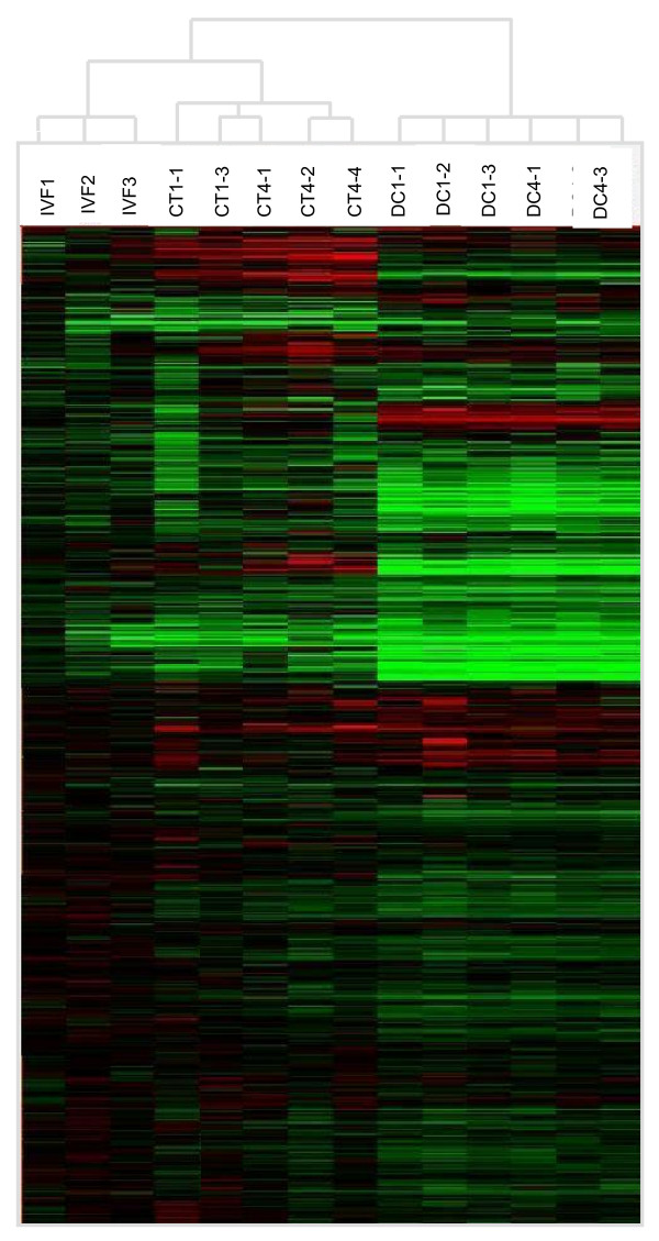
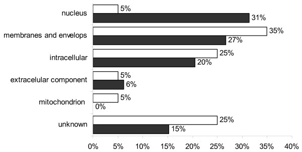
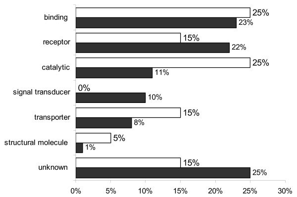
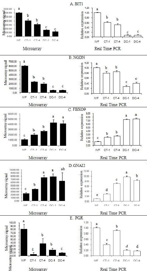
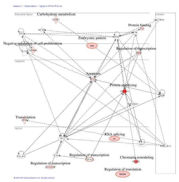
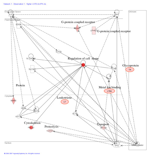
Results: Bovine clones from up to four generations of successive cloning were produced by chromatin transfer (CT). Using Affymetrix bovine microarrays we determined that the transcriptomes of blastocysts derived from the first and the fourth rounds of cloning (CT1 and CT4 respectively) have undergone an extensive reprogramming and were more similar to blastocysts derived from in vitro fertilization (IVF) than to the donor cells used for the first and the fourth rounds of chromatin transfer (DC1 and DC4 respectively). However a set of transcripts in the cloned embryos showed a misregulated pattern when compared to IVF embryos. Among the genes consistently upregulated in both CT groups compared to the IVF embryos were genes involved in regulation of cytoskeleton and cell shape. Among the genes consistently upregulated in IVF embryos compared to both CT groups were genes involved in chromatin remodelling and stress coping.

Conclusion: The present study provides a data set that could contribute in our understanding of epigenetic errors in somatic cell chromatin transfer. Identifying "cumulative errors" after serial cloning could reveal some of the epigenetic reprogramming blocks shedding light on the reprogramming process, important for both basic and applied research.


Figures

Figure 1
Figure 1
Agilent bioanalyzer gel-like image of total RNA. The image shows a total RNA gel like-image produced by the Bioanalyzer. (Ten out of the 15 samples used in the microarray experiment are shown since no more than 11 samples can be run at one time). Lane L: Size markers. Lanes 1 and 2: total RNA from 106 donor cells used for the first round of embryonic cloning. Lanes 3 and 4: total RNA from 106 donor cell used for the fourth round of cloning. Lanes 5 and 6: total RNA from a pool of 3 In Vitro Produced embryos. Lanes 7 and 8: total RNA from a pool of 3 embryos produced by the first round of chromatin transfer. Lanes 9 and 10: total RNA from a pool of 3 embryos produced by the fourth round of chromatin transfer. The 28S and 18s distinctive ribosomal RNA bands are observed for all samples.
Figure 2
Figure 2
Hierarchical clustering of microarray hybridizations. Cluster analysis of hybridizations and genes performed using GeneTraffic UNO (Iobion Informatics LLC). All donor cells were clustered in one group, while all the embryos were clustered in a second group. The embryos clearly separate into two groups: a group containing the IVF embryos and a group containing the chromatin transfer embryos.
Figure 3
Figure 3
GoSlimViewer graph of Cellular Component over-represented terms in IVF and CT embryos. Sub-cellular locations of gene products found at high levels in both IVF blastocysts (solid bars) and both groups of CT blastocysts (open bars). The proportion of genes present in the nucleus was higher in IVF embryos (31%) compared to CT embryos (5%). There were more membrane and intracellular genes in CT embryos compared to IVF embryos.
Figure 4
Figure 4
GoSlimViewer graph of Biological Process over-represented terms in IVF and CT embryos. Biological processes of gene products found at high levels in both IVF blastocysts (solid bars) and CT blastocysts (open bars). No genes involved in development were upregulated in CT blastocysts compared to IVF blastocysts, for which 11% of the genes were involved in development. Conversely a greater proportion of metabolism genes were overrepresented in CT embryos compared to IVF embryos.
Figure 5
Figure 5
GoSlimViewer graph of Molecular Function over-represented terms in IVF and CT embryos. Molecular functions of gene products found at high levels in IVF blastocysts (solid bars) and CT blastocysts (open bars). Genes with receptor function were higher in IVF blastocysts, while genes with catalytic, signal transduction and transporter functions were overrepresented in CT blastocysts.
Figure 6
Figure 6
Real Time PCR gene expression analysis. Validation of gene expression patterns from the microarray analysis (black bars) by relative quantification through Real time PCR (open bars). A. Validation of gene expression patterns of PLAC8. B. Validation of gene expression patterns of HSPA1. C. Validation of gene expression patterns of HMGN3. D. Validation of gene expression patterns of DNMT3a. E. Validation of gene expression patterns of DNMT3b. F. Validation of gene expression patterns of IGF2R.
Figure 7
Figure 7
Real Time PCR gene expression analysis. Validation of gene expression patterns from the microarray analysis (black bars) by relative quantification through Real time PCR (open bars). A. Validation of gene expression patterns of BIT1. B. Validation of gene expression patterns of NGDN. C. Validation of gene expression patterns of FBXO9. D. Validation of gene expression patterns of GNAI2. E. Validation of gene expression patterns of PGR. Real time PCR units indicate relative expression to the internal standard GAPDH. Different letters on top of each bar indicate significant differences in expression (P < 0.01).
Figure 8
Figure 8
Real Time PCR gene expression analysis in bovine donor cells. Gene expression analysis of PALLD, NFYA, GATM and Taspase1 in donor cells lines derived from 0 rounds of cloning (DC0) first round of cloning (DC1), second round of cloning (DC2), fourth round of cloning (DC4), and fifth round of cloning (DC5). Units indicate relative expression to the internal standards GAPDH and 18S rRNA. Different letters indicate significant differences in expression between different donor cell lines (P < 0.01).
Figure 9
Figure 9
Display of genes with high expression in IVF embryos. Data modelling of genes with high expression in IVF embryos compared to cloned embryos. The top networks in the pathway include cellular growth and proliferation, embryonic development, cellular assembly and organization, cellular death and response to stress and cancer.
Figure 10
Figure 10
Display of genes with high expression in CT embryos. Data modelling of genes with higher expression in CT embryos compared to IVF embryos. The top networks in the pathway include cellular morphology, cellular development, cell signalling and metabolism.

References

    1. Memili E, First NL. Zygotic and embryonic gene expression in cow: a review of timing and mechanisms of early gene expression as compared with other species. Zygote. 2000;8:87–96. - PubMed
    1. Whitworth K, Springer GK, Forrester LJ, Spollen WG, Ries J, Lamberson WR, Bivens N, Murphy CN, Mathialagan N, Green JA, et al. Developmental expression of 2489 gene clusters during pig embryogenesis: an expressed sequence tag project. Biol Reprod. 2004;71:1230–1243. - PubMed
    1. Latham KE, Schultz RM. Embryonic genome activation. Front Biosci. 2001;6:D748–759. - PubMed
    1. Han YM, Kang YK, Koo DB, Lee KK. Nuclear reprogramming of cloned embryos produced in vitro. Theriogenology. 2003;59:33–44. - PubMed
    1. Vajta G, Gjerris M. Science and technology of farm animal cloning: state of the art. Anim Reprod Sci. 2006;92:211–230. - PubMed

Publication types